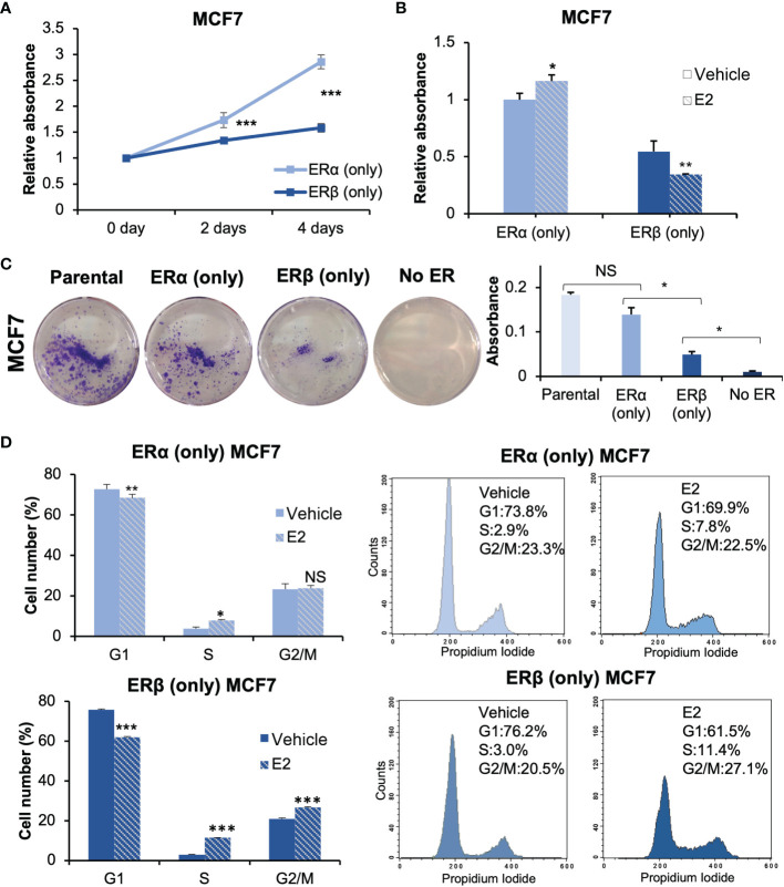
Figure 2.
ERα and ERβ impact cell proliferation differently. (A) Cell proliferation of ERα (only) or ERβ (only) MCF7 cells was measured using WST-1 assay. Cells were grown in full-serum medium and measured at day 0, 2, and 4. Absorbance at day 0 was used for normalization. (B) The cell lines were pre-cultured under non-estrogenic and serum-starved conditions, followed by E2 or vehicle treatment and measured by WST-1 assay at day 4. Absorbance of ERα (only) MCF7 cells with vehicle stimulation was used for normalization. (C) For clonogenic assay, the cells were cultured in full-serum medium for 8 days. Extracted crystal violet was used for quantification (right). (D) Flow cytometry analysis of cell cycle progression of ERα (only) or ERβ (only) MCF7 cells (right) and corresponding quantitation of cell cycle distribution (G1, S and G2/M, left). Cells were grown in 2.5% DCC-FBS medium for 72 h, followed by treatment of E2 or vehicle for 24 h. Data is illustrated as means ± SD (n=3). A, B, D were analyzed using two-way ANOVA followed by Bonferroni test; C was analyzed using one-way ANOVA. *P < 0.05, **P < 0.01, ***P < 0.001, NS, not significant.

