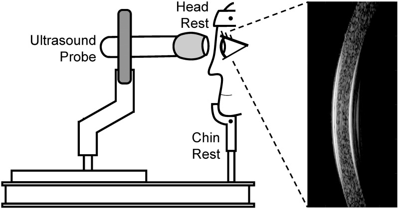
Fig 1. OPE measurement setup showing a subject sitting in front of an anti-vibration table with head secured into a chin-and-head rest mounted on the table.
The ultrasound probe is mounted on a holder whose XYZ position can be adjusted by an operator. Ultrasound B-mode images and RF data of the cornea are collected along the nasal-temporal cross-section centered at the cornea apex.

