
FIGURE 2.
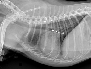
Right lateral thoracic radiographic image of a CKCS demonstrating the radiographic measurements of vertebral left atrial size (VLAS) perfomed in this study (kVp 75, mAs 2.5). A line was drawn from the central and ventral border of the carina to the caudal most border of the left atrium, where it intersected with the dorsal border of the caudal vena cava (#). This line was indexed to the thoracic vertebral bodies starting at the cranial edge of T4 (*) and summed (1.7 vertebrae in this example)