
FIGURE 6.
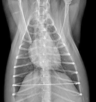
Dorso‐ventral thoracic radiographic image of a CKCS demonstrating the radiographic measurements of thoracic width measured as the distance between medial borders of eighth rib at their most lateral curvatures in dorso‐ventral recumbency (kVp 75, mAs 2.5)