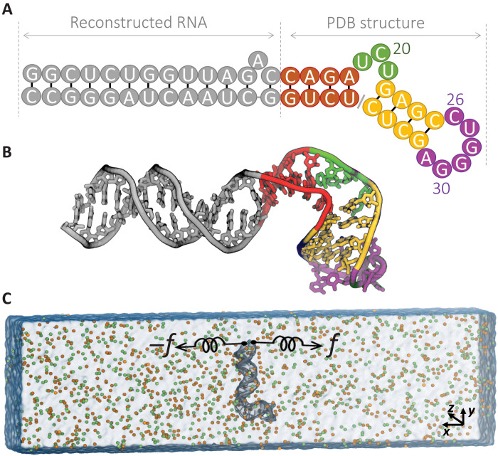
Fig. 1. Schematic of molecular simulation setup for HIV-1 TAR hairpin.
(A) RNA sequence. (B) The structure of the HIV-1 TAR hairpin under study consists of an extended stem (gray), connected to lower stem (red) with apical loop (green), followed by upper stem (yellow) that is bridged by bulge region (purple). (C) Setup for the MD simulation in 400 mM KCl, K+ (orange) and Cl− (green). The RNA hairpin (gray) is aligned perpendicular to the x axis, and the terminus nucleotides are pulled.

