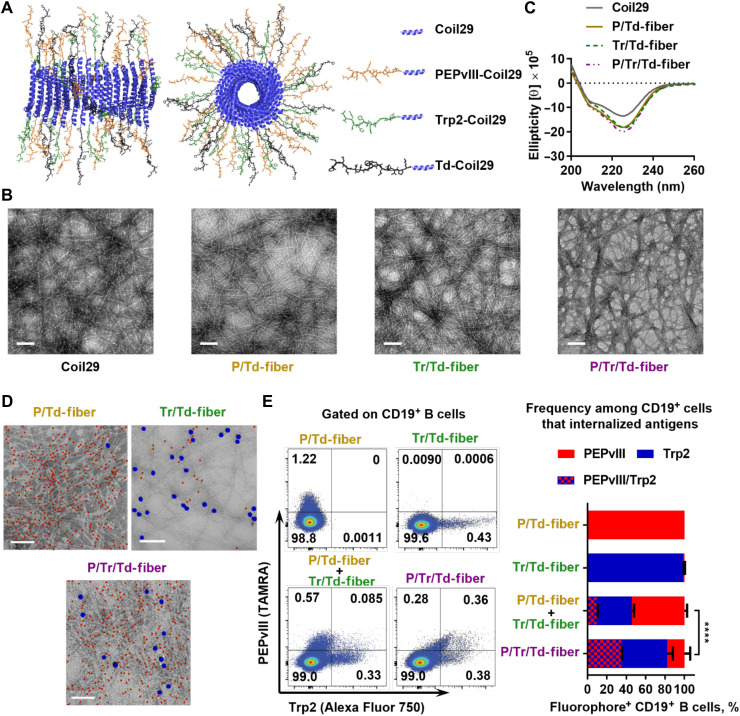
Fig. 1. Co-assembled Coil29 peptides exhibit fibril morphologies and deliver both B cell and T cell peptide epitopes simultaneously.
(A) Schematics of α-helical Coil29 nanofiber carrying multiple epitopes, drawn using Protein Data Bank (PDB) structures for Coil29 (PDB ID 3J89). Left, side view; right, axial view. (B) All assemblies exhibited similar α-helical secondary structures by CD, consistent with unmodified Coil29. (C) Multiepitope Coil29 nanofibers exhibited fibrillar morphologies similar to unmodified Coil29 by TEM. Scale bar, 100 nm. (D) Representative immunogold-labeled TEM images indicate that co-assembled nanofibers presented both Trp2 (5 nm) and PEPvIII (10 nm) epitopes Scale bar, 100 nm. Five-nanometer particles (PEPvIII labeling) are false-colored red, and 10-nm particles (Trp2 labeling) are false-colored blue for clarity. (E) Uptake of assembled fluorophore-labeled peptides (TAMRA-PEPvIII-Coil29 and Alexa Fluor 750–Trp2–Coil29) by CD19+ B cells 4 hours after intraperitoneal administration of either single fluorophore-labeled nanofibers (PE or Alexa Fluor 750) or two fluorophore-labeled co-assembled nanofibers (PE/Alexa Fluor 750) confirmed co-assembly between Trp2-Coil29 and PEPvIII-Coil29 peptides. Left, representative flow histograms; right, percentage of B cells internalizing either or both fluorophores among the fluorophore-positive B cells. (****P < 0.0001 by χ2 test, N = 3).

