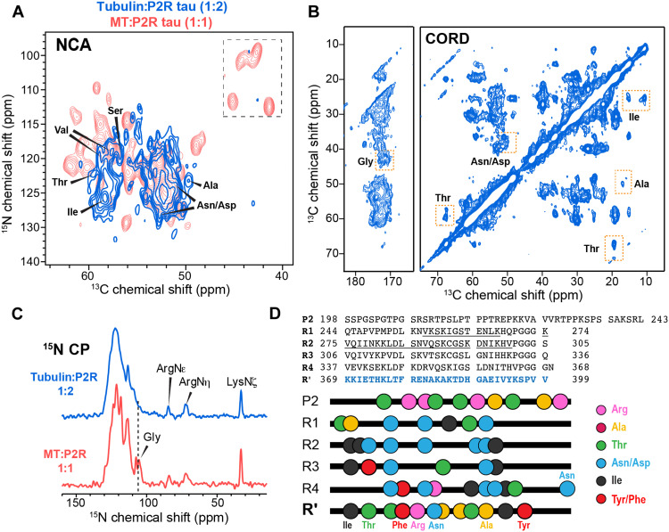
Fig. 3. R′ binds both dynamically unstable MTs.
(A) 2D NCα spectrum of tubulin-tau (1:2) coassembly (blue), overlaid with the spectrum of taxol-stabilized MT-P2R tau (1:1) complex (red). (B) 2D CC correlation spectrum of tubulin-tau coassembly. Joint analysis of the two 2D spectra allows assignment of the immobilized residues. Thr, Asn, Asp, Ala, and Ile signals are observed. (C) 15N CP spectra of P2R tau bound to unstable (top) and stabilized (bottom) microtubules. Arginine side-chain signals are detected in both spectra, indicating that some Arg side chains are immobilized by the MTs. The Gly 15N signals are broadened in the coassembled sample relative to the taxol-stabilized sample. (D) Amino acid sequence of P2R tau and the location of key residue types that identify the immobilized segment in the tubulin:tau coassembly. R′ is the only segment that contains all residue types (Ala, Arg, Thr, Ile, Tyr/Phe, and Asp/Asn) in the dipolar spectra. The cryo-EM reported MT-bound R1 and R2 segments in chimeric tau constructs are underlined.

