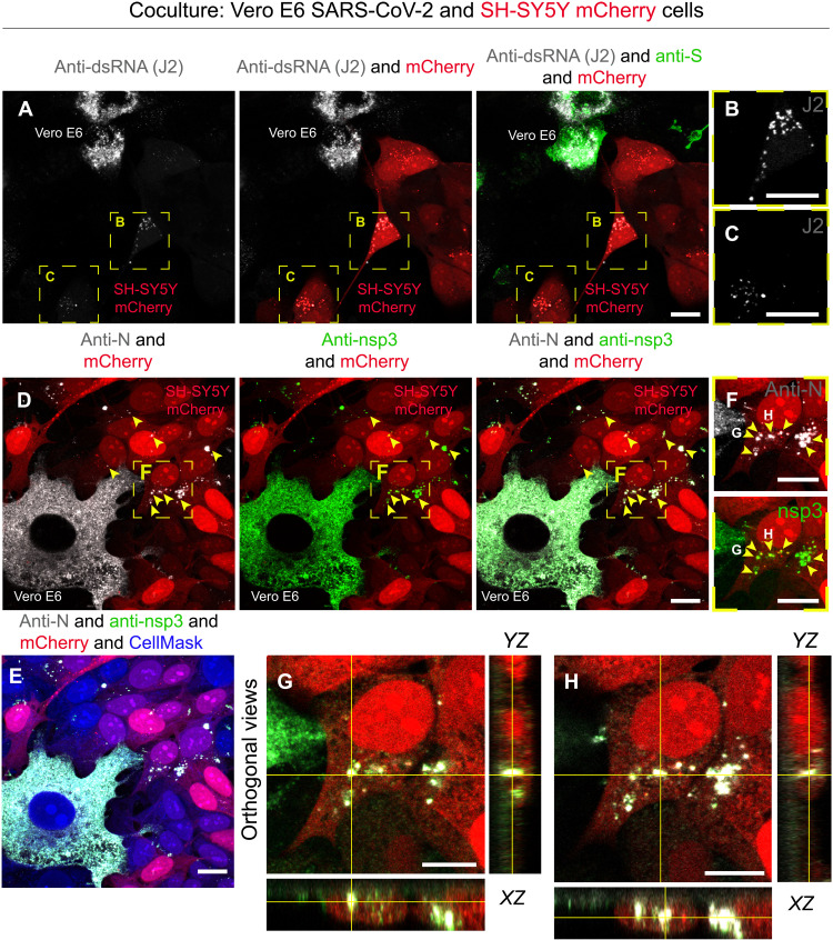
Fig. 2. Anti-dsRNA antibody J2 and the nonstructural protein 3 are detected in SH-SY5Y cells cocultured with SARS-CoV-2–infected Vero E6 cells.
(A) SARS-CoV-2–infected Vero E6 cells (donor cells) were cocultured for 48 hours with SH-SY5Y mCherry acceptor cells. Confocal micrographs showing the staining with J2 antibody are used to detect dsRNA, and an anti-S antibody is used to detect SARS-CoV-2 particles. (B and C) Enlargement of the yellow dashed squares in (A). (D to H) Confocal micrographs showing 48 hours of coculture of SARS-CoV-2–infected Vero E6 cells (donor) and SH-SY5Y mCherry cells (acceptor) stained using anti–nonstructural protein 3 (nsp3) and anti-N antibodies. (E) Confocal micrographs showing cellular cytoplasm labeled with CellMask Blue. (F) Enlargement of the yellow dashed square in (D) showing puncta positive for both anti-nsp3 and anti-N in acceptor cells. (G and H) Confocal micrographs representing the orthogonal views of (F) showing anti-nsp3 and anti-N puncta in the cytoplasm of acceptor cells. Yellow arrowheads indicate anti-nsp3 and anti-N signal in acceptor cells. Scale bars, 10 μm.

