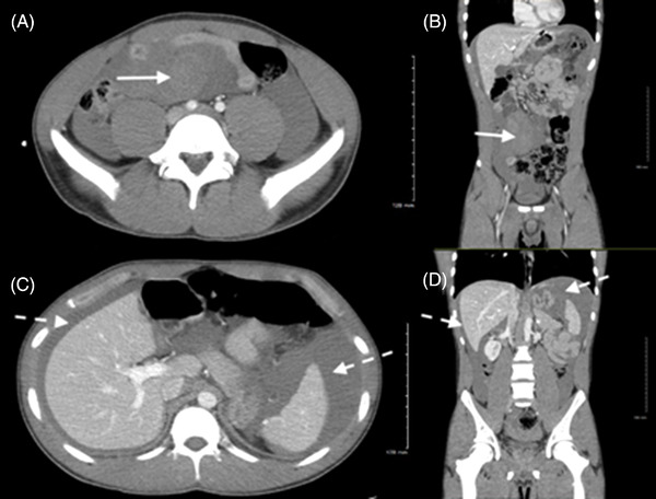
FIGURE 2.

Computed tomography of the abdomen/pelvis demonstrating a localized mesenteric hematoma (solid arrow) of the right mid lower abdomen in axial (A) and coronal (B) planes. Computed tomography of the abdomen/pelvis demonstrating a moderate amount of fluid and blood (dotted arrows) within the peritoneal cavity in axial (C) and coronal (D) planes
