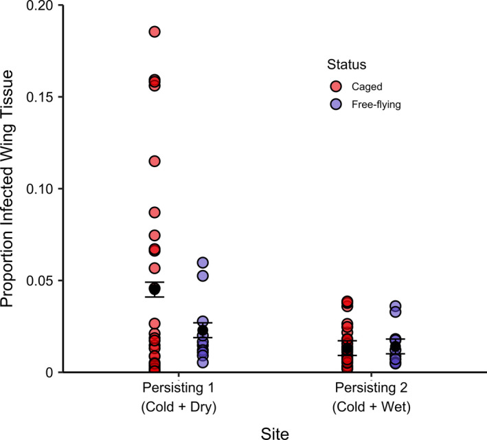
FIGURE 6.

Proportion of infected wing tissue as indicated by orange fluorescence on caged and free‐flying bats in each of the two persisting sites (Meteyer et al., 2009; Turner et al., 2014). Point colours represent caging status: caged = red, free‐flying = blue. Black points and error bars represent model estimates ± standard error. Free‐flying bats were opportunistically captured at the end of hibernation at experiment termination. In Persisting 1 (Cold + Dry), where disease severity in caged bats was high, caged bats had significantly higher levels of tissue invasion compared to free‐flying bats within the same site. However, this difference was not detected at Persisting 2 (Cold + Wet). This suggests that little brown bats in the cold and dry persisting site do not roost in dry microclimates for the entirety of hibernation, but rather move roosting locations periodically, as has been observed prior to the WNS epidemic. Moving amongst a variety of microclimates in this site may allow this persisting colony to mitigate disease severity
