. 2022 Jul 7;11:e71437. doi: 10.7554/eLife.71437

Figure 5. Thin localizes in close proximity to Dysbindin.

(A) Confocal maximum intensity projection of a representative neuromuscular junction (NMJ) branch (muscle 6–7) after presynaptic coexpression (elavc155-Gal4) of venus-tagged Dysbindin (UAS-venus-Dysbindin, ‘Dysbvenus’, green) and mCherry-tagged Thin (UAS-mCherry-thin, ‘ThinmCherry’, magenta) detected with anti-GFP and anti-DsRed, respectively. (B) Single plane of the synaptic bouton highlighted by the white square in (A) with corresponding line profile (right). The yellow line demarks the location of the line profile. (C) gSTED image of the synaptic bouton shown in (B) with corresponding line profile (right). Scale bar, A: 5 µm; B, C: 2 µm. Note the partial overlap between ThinmCherry and Dysbindinvenus at confocal and STED resolution. (D) Left: Schematic of nearest-neighbor (NND) analysis between ThinmCherry and Dysbindinvenus puncta at STED resolution. Right: ThinmCherry puncta (‘+’, maximum locations, see Materials and methods) and the NNDs and locations of Dysbindinvenus puncta (color code denotes NND) of a representative bouton. (E) Histogram of mean ThinmCherry − Dysbindinvenus NND per bouton of the recorded gSTED data (blue), or after randomized punctum distribution (gray, see Materials and methods). N = 10 NMJs, average n = 13 boutons per NMJ for data and simulations. Observed vs. randomized NNDs, p < 0.001; Student’s t-test.

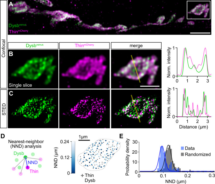
Figure 5—source data 1. Related to Figure 5.

Figure 5.

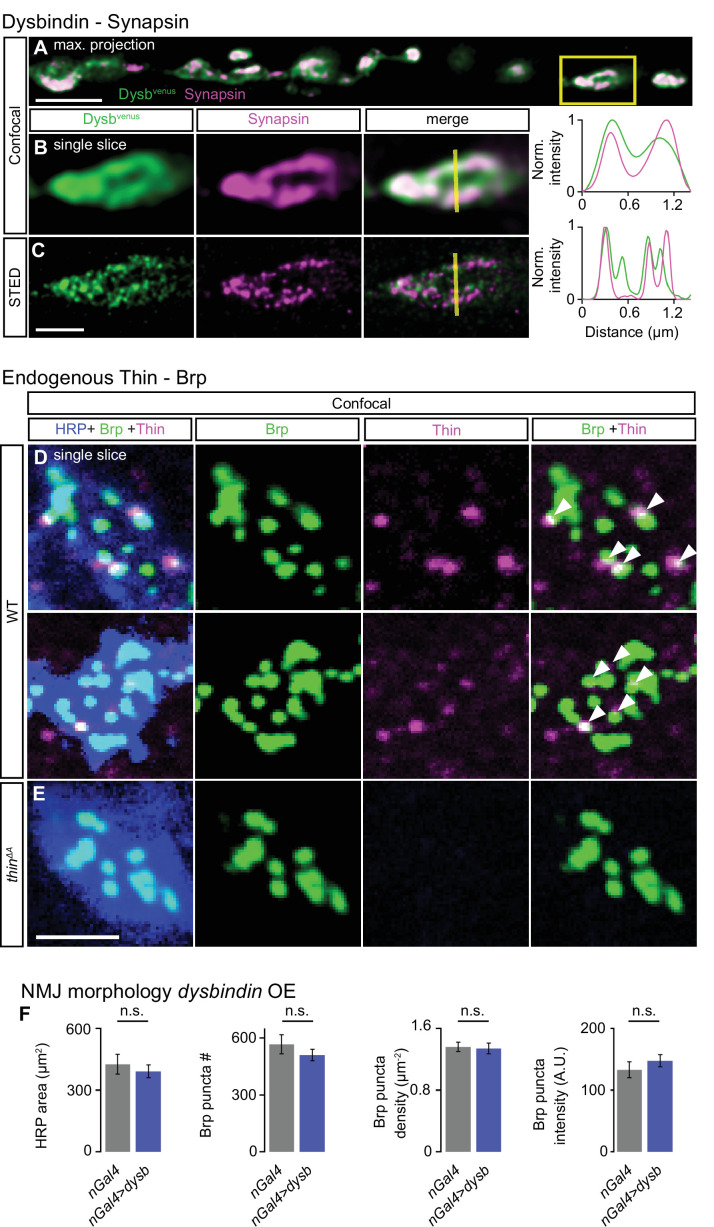
Figure 5—figure supplement 1. Dysbindin and Synapsin distribute in the periphery of synaptic boutons, endogenous Thin localizes close to Brp, and presynaptic dysbindin overexpression does not affect neuromuscular junction (NMJ) morphology.

Figure 5—figure supplement 1.

(A) Confocal maximum intensity projection of a representative NMJ branch (muscle 6–7) after presynaptic expression (elavc155-Gal4) of venus-tagged Dysbindin (UAS-venus-dysbindin, ‘Dysbvenus’) stained with anti-GFP (green, ‘Dysbvenus’) and anti-Synapsin (magenta, ‘Synapsin’). (B) Single slice of the synaptic bouton highlighted by the yellow square in (A) with corresponding line profile (right). The yellow line demarks the location of the line profile. (C) gSTED image of the synaptic bouton shown in (B) with corresponding line profile (right). Note the partial overlap between Dysbvenus and the synaptic vesicle marker Synapsin at confocal and STED resolution. Scale bar, A: 4 µm; B, C: 1 µm. (D) Confocal single slice of two representative wild-type (WT) NMJ boutons (muscle 6) stained with the neuronal membrane marker anti-HRP (‘HRP’, blue), the active-zone marker Bruchpilot (anti-Brpnc82, ‘Brp’, green), and anti-Thin (‘LaBeau-DiMenna et al., 2012, magenta). (E) Same staining as in (D) for a thinΔA mutant NMJ. Note that some Thin puncta localize in close proximity to Brp within WT boutons (white arrowheads, D, right), suggesting presynaptic Thin puncta in the vicinity of AZs. A quantitative analysis of the relationship between anti-Thin and presynaptic markers could not be realized because of postsynaptic anti-Thin puncta in the muscle cell (not shown, LaBeau-DiMenna et al., 2012). Little to no anti-Thin signal was detected at thinΔA mutant NMJs (E). Scale bar, 1 µm. (F) Mean HRP area per muscle 6/7 NMJ (‘HRP area’), Brp puncta number per NMJ (‘Brp puncta #’), Brp puncta number/HRP area per NMJ (‘Brp density’), and Brp puncta fluorescence intensity of control (elavc155-Gal4/+, ‘nGal4’, gray) and after presynaptic expression of venus-tagged Dysbindin in WT (elavc155-Gal4>UAS-venus-dysbindin, ‘nGal4>dysb’, blue). Note that all morphological parameters were largely unchanged after presynaptic dysbindin overexpression, implying no major changes in NMJ morphology. Mean ± standard error of the mean (SEM); nGal4: n = 9, nGal4>Dysb: n = 11; n.s.: not significant; Student’s t-test.
Figure 5—figure supplement 1—source data 1. Related to Figure 5—figure supplement 1F.
Dysbindin and Synapsin distribute in the periphery of synaptic boutons, endogenous Thin localizes close to Brp, and presynaptic dysbindin overexpression does not affect neuromuscular junction (NMJ) morphology.
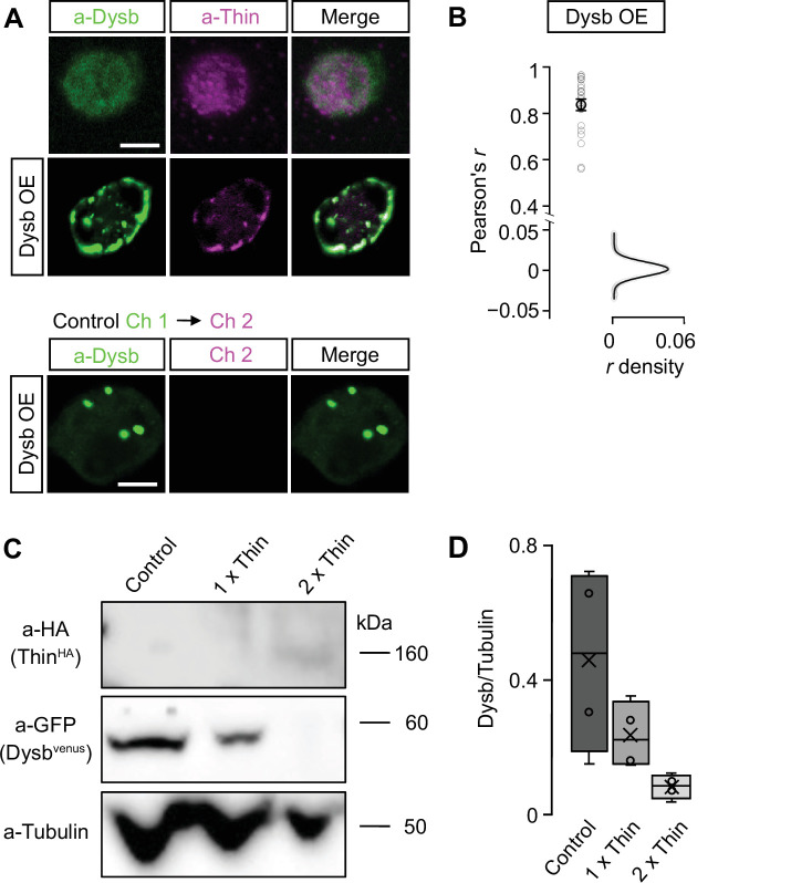
Figure 5—figure supplement 2. Thin localizes in close proximity to Dysbindin and Thin degrades Dysbindin in Drosophila S2 cells.

Figure 5—figure supplement 2.

(A) Confocal images (single planes) of Drosophila S2 cells stained with anti-Dysbindin (green) and anti-Thin (magenta) under control conditions (top) and after dysbindin overexpression (UAS-venus-Dysbindin, ‘Dysb OE’, middle). Note the redistribution of Thin upon dysbindin expression resulting in overlapping Thin and Dysbindin fluorescence. Bottom: Excitation of channel 1 (anti-Dysbindin) did not produce significant fluorescence in channel 2 (anti-Thin), implying no major crosstalk between the two channels. (B) Pearson’s correlation coefficient (r) between anti-Dysbindin and anti-Thin fluorescence intensities per pixel after dysbindin overexpression (‘Dsyb OE’) (n = 22). The r density (with Gaussian fit) was obtained from simulated Thin and Dysbindin localizations after sampling random point spread function-sized chunks of the data (see Materials and methods, n = 22). The average observed r = 0.84 is significantly higher than expected from random Thin and Dysbindin localizations. (C) Representative western Blot of S2 cells transfected for 72 hr with constant levels of pUAS-venus-Dysbindin (‘Dysbindinvenus’) and the indicated relative cDNA concentrations of pUAS-HA-thin (‘ThinHA’) normalized to pUAS-venus-dysbindin (‘1×’, ‘2×’). Dysbindinvenus and ThinHA were detected with anti-GFP (‘a-GFP’) and anti-HA (‘a-HA’), respectively. Anti-Tubulin (‘a-Tubulin’) served as a loading control. (D) Quantification of Dysbindinvenus/Tubulin fluorescence intensity at different relative ThinHA concentrations (‘1×’, ‘2×’) relative to Dysbindinvenus (n = 4). Note the decrease in Dysbindinvenus/Tubulin upon thin overexpression. Scale bar, A: 5 µm; B, C: 2 µm.
Figure 5—figure supplement 2—source data 1. Related to Figure 5—figure supplement 2.
Thin localizes in close proximity to Dysbindin and Thin degrades Dysbindin in Drosophila S2 cells.