FIGURE 5.

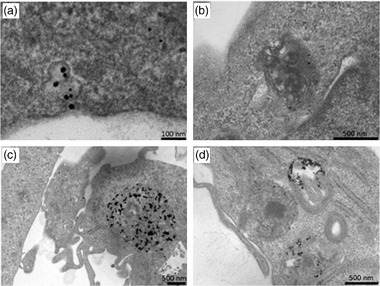
Transmission electron microscopy images of MDA‐MB‐231 cells treated with 200 μg/mL AuNPs 15 nm for 6 h (a, b) or for 24 h (c, d). 60‐nm thick cell sections.

Transmission electron microscopy images of MDA‐MB‐231 cells treated with 200 μg/mL AuNPs 15 nm for 6 h (a, b) or for 24 h (c, d). 60‐nm thick cell sections.