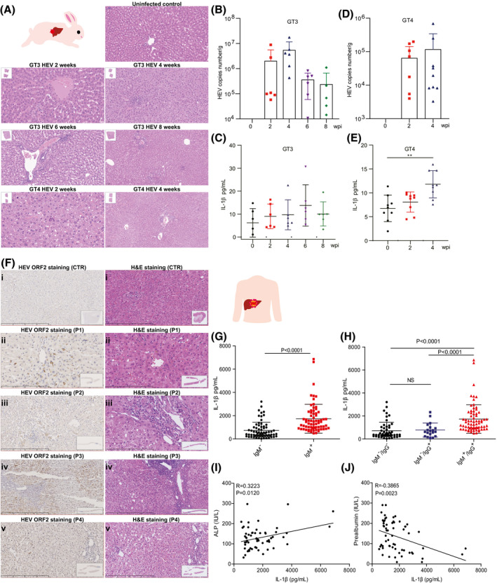
FIGURE 1.

HEV infection significantly elevates IL‐1β secretion, the hallmark of inflammasome activation, in rabbits and patients. (A) Rabbits were inoculated with GT3 and ‐4 HEV. Histology showing liver inflammation in infected, but not in the control, animals. (B,C) Shedding of GT3 HEV into feces and secretion of IL‐1β into blood were measured at 0, 2, 4, 6, or 8 wpi. (D,E) Shedding of GT4 HEV and secretion of IL‐1β were measured at 0, 2, and 4 wpi. (F) i is control tissue, which was derived from a hepatic hemangioma patient without HEV infection. i showed no clear liver injury. ii, iii, iv, and v tissues were from 4 different HEV‐infected patients, respectively, showing different histological/pathological features. ii shows apparent cholestasis in hepatic bile capillary with pigment‐containing granulocytophagous cells. iii shows infiltration of inflammatory cells in the portal area. iv shows an enlarged portal area with abundant inflammatory cells with a tendency of fibrosis. v shows some punctate necrosis and infiltration of a small amount of inflammatory cells in the portal area. (G) IL‐1β levels in serum of patients (IgM positive; n = 70) and healthy persons (IgM negative; n = 70) were determined by ELISA. (H) IL‐1β levels in serum of patients (IgM positive; n = 70) and in healthy controls who never encountered HEV (IgM−/IgG−; n = 19) or who were historically exposed to HEV (IgM−/IgG+; n = 51). Correlations between ALP (I) or prealbumin (J) and IL‐1β were analyzed. Data are means ± SD. *p < 0.05; **p < 0.01; ***p < 0.001; ****p < 0.0001. Abbreviations: CTR, control; NS, not significant (Mann‐Whitney U test); P, patient
