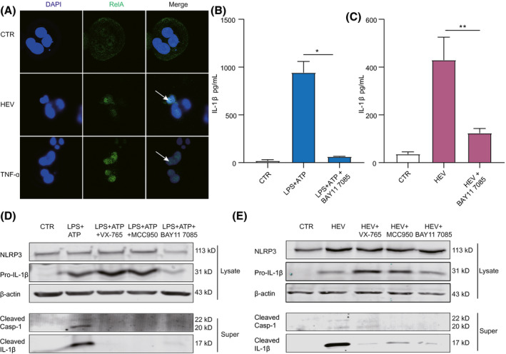
FIGURE 4.

NF‐κB signaling activation by HEV is essential for NLRP3 inflammasome response. (A) THP‐1 macrophages were inoculated with HEV or treated with TNF‐ɑ (100 ng/mL) for 24 hours. Subcellular localizations of NF‐κB (green) and the nucleus marker, DAPI (blue), were examined under confocal microscopy. (B) THP‐1 macrophages were pretreated with 10 µM of NF‐κB inhibitor (BAY11 7085) for 2 hours and then treated with LPS (400 ng/ml) or LPS (400 ng/mL) plus 10 µM of NF‐κB inhibitor (BAY11 7085) for 6 hours, followed by ATP (5 mM) for 40 minutes. IL‐1β level in supernatant was quantified by ELISA (n = 4). (C) THP‐1 macrophages were pretreated with 10 µM of NF‐κB inhibitor (BAY11 7085) for 2 hours and then inoculated with HEV plus 10 µM of NF‐κB inhibitor (BAY11 7085) for 24 hours. IL‐1β level in supernatant was quantified by ELISA (n = 4). (D) Mature IL‐1β and cleaved Casp‐1 in supernatant or pro‐IL‐1β and NLRP3 in lysates were determined after LPS plus NF‐κB inhibitor treatment by western blotting. (E) Mature IL‐1β and cleaved Casp‐1 in supernatant or pro‐IL‐1β and NLRP3 in lysates were determined after HEV infection plus NF‐κB inhibitor by western blotting. NF‐κB activation is marked by an arrow. Data were normalized to the control (CTR; set as 1). Data are means ± SD. *p < 0.05; **p < 0.01; ***p < 0.001; Abbreviations: NS, not significant (Mann‐Whitney U test); Super, supernatant
