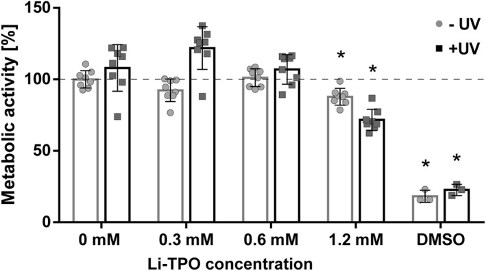
FIGURE 1.

Metabolic activity of human articular chondrocytes (P3) exposed to different concentrations of lithium phenyl‐2,4,6‐trimethyl‐benzoylphosphinate (Li‐TPO) with and without exposure to UV light. Metabolic activity was measured by resazurin‐based Presto Blue staining after 2 h of Li‐TPO exposure followed by 24 h of incubation. Presto Blue fluorescence of cells treated with different Li‐TPO concentrations is shown as mean percentage ± standard deviation compared to control (no Li‐TPO). n = 8 for each group. * highlights significant differences (p < 0.001) compared to control ‐UV. There was no difference between the 0.6 mM Li‐TPO and control groups. (Dimethylsulfoxide [DMSO] = negative control, n = 3)
