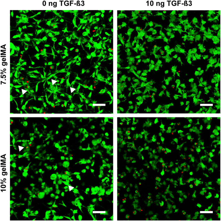
FIGURE 3.

Cell morphology of human articular chondrocytes P3 after 3 weeks of encapsulation in soft (7.5%) and stiff (10%) gelatin methacryloyl (gelMA) with and without TGF‐ß3. Without TGF‐ß3, cell morphology was highly heterogenous in both soft as well as stiff gelMA. Although spindle‐shaped (arrow heads) and round cells were found in both stiffnesses, the round cell morphology (chondrocyte like) was favored in the stiffer gelMA, whereas the spindle‐like morphology (fibroblast like) was dominant in the softer gelMA. In the 10 ng TGF‐ß3 group both stiffnesses contained a rather homogenous cell population of roundish or polygonal cells with little cell processes. Scale bar: 50 µm
