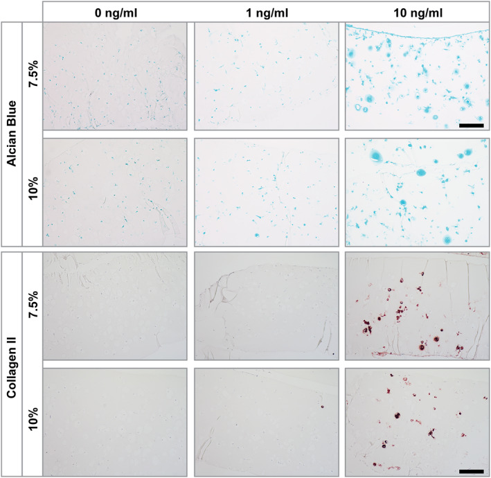
FIGURE 5.

Alcian blue and collagen type II stainings of human articular chondrocytes (hAC) (P3, Donor 3) encapsulated in soft (7.5%) and stiff (10%) gelatin methacryloyl (gelMA) after 21 days in culture containing different concentrations of TGF‐ß3 (0, 1 and 10 ng/mL). Culture with 0 ng/mL and 1 ng/mL TGF‐ß3 displayed a lack of collagen type II staining. Glycosaminoglycans staining was absent in 0 ng/mL TGF‐β3 cultures but showed slight staining at 1 ng/mL TGF‐β3. HAC cultures within 10 ng/mL TGF‐ß3 both stainings clearly showed positive cells. The staining was located in close proximity to the cells and within the gelMA matrix. Scale bar: 100 µm
