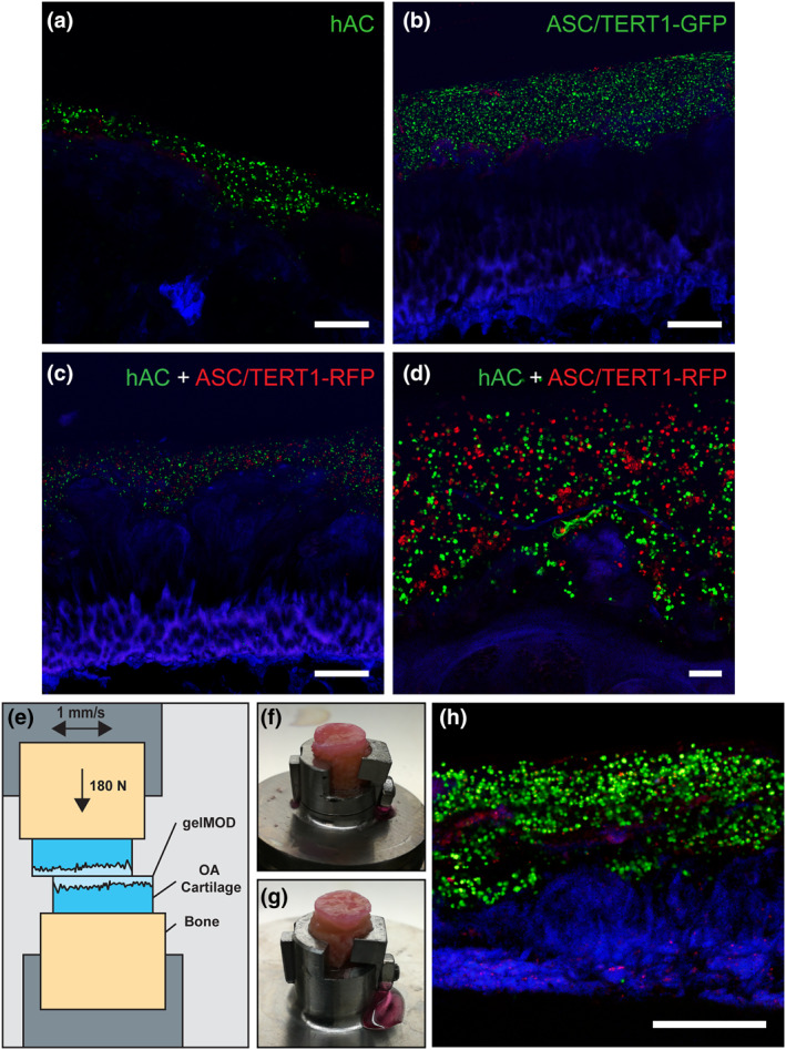
FIGURE 6.

Superficially damaged osteoarthritis (OA) cartilage coated with cell‐loaded gelatin methacryloyl (gelMA) (10%) after 1 day of cultivation and after simulation of human gait. (a) gelMA loaded with human articular chondrocytes (hAC) (DiO, green) and stained for dead cells (ethidium homodimer 1; red) on cartilage (autofluorescence; blue). (b) gelMA loaded with ASC/TERT1‐GFP (GFP‐transduced; green) and stained for dead cells (ethidium homodimer 1; red) (c) Overview and (d) detail of co‐culture of hAC (green) and ASC/TERT1‐mCherry (red). Scale bar: (a–c) 500 µm and (d) 100 µm. (e) Schematic of the experimental setup for the mechanical simulation of human gait. Osteoarthritic specimens were coated with cell loaded (hAC‐DiO) gelMA (10%) and exposed to mechanical stress to simulate human gait. The gelMA layer stayed intact in both specimens: (f) lower specimen of the measurement, (g) upper specimen. (h) Live/Dead staining of a cross‐section of the lower specimen showing living cells (green), dead cells (red) and cartilage (blue). Scale bar: 500 µm
