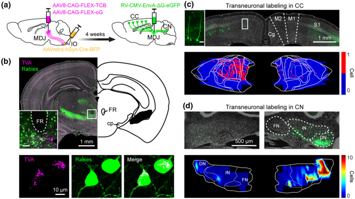
FIGURE 6.

Monosynaptic rabies tracing revealing CC and CN inputs on the olivary‐projecting MDJ neurons. (a) Schematic of transneuronal viral‐tracing strategy (n = 4 mice). (b) Coronal sections of a representative mouse at the midbrain level (upper, inset suggesting MDJ, inset scale bar = 100 µm), showing rabies primary labeling (lower, TVA and rabies co‐labeled) and local secondary labeling. (c) Coronal section (upper) and plotting map (lower) of transneuronal rabies labeling in layer‐5 pyramidal cells (inset, scale bar = 50 µm) of motor cortex. (d) Same as (c), but for labeling in the CN. DN, dentate nucleus; FN, fastigial nucleus; IN, interposed nucleus
