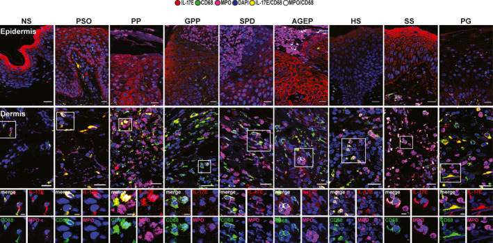
Figure 1.

Interleukin (IL)‐17E immunofluorescence staining (mouse anti‐IL‐17E, red) in macrophages (rabbit anti‐CD68, green) and neutrophils [goat anti‐myeloperoxidase (MPO), pink] in the epidermis (first row) and dermis (second row and higher magnification). One representative cropped image is shown. Original magnification × 40; scale bar 20 µm (higher magnification scale bar 5 µm). AGEP, acute generalized exanthematous pustulosis; GPP, generalized pustular psoriasis; HS, hidradenitis suppurativa; NS, normal skin; PG, pyoderma gangrenosum; PP, paradoxical psoriasis; PsO, chronic plaque‐type psoriasis; SPD, subcorneal pustular dermatosis of Sneddon–Wilkinson; SS, Sweet syndrome.
