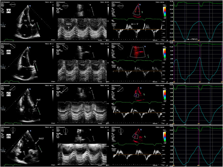
FIGURE 2.

Echocardiographic images of the four multi‐plane right ventricular (RV) views (A‐D) with corresponding quantitative functional parameters of the respective free wall segments (L‐R panels). (A) – Focused four chamber view (0°), lateral wall; (B) – coronary sinus view (+40°), anterior wall; (C) – aortic view (‐40°), inferior wall; (D) – coronal view (‐90°), inferior wall and RVOT anterior wall. Second panel (center left): tricuspid annular plane systolic excursion (TAPSE); third panel, (center right): tricuspid annular peak systolic velocity (RV‐S’); fourth panel (far right): RV wall longitudinal strain (RV‐LS). LV ‐ left ventricle; CS ‐ coronary sinus; AoV ‐ aortic valve; RVOT ‐ right ventricular outflow tract
