
FIGURE 4.
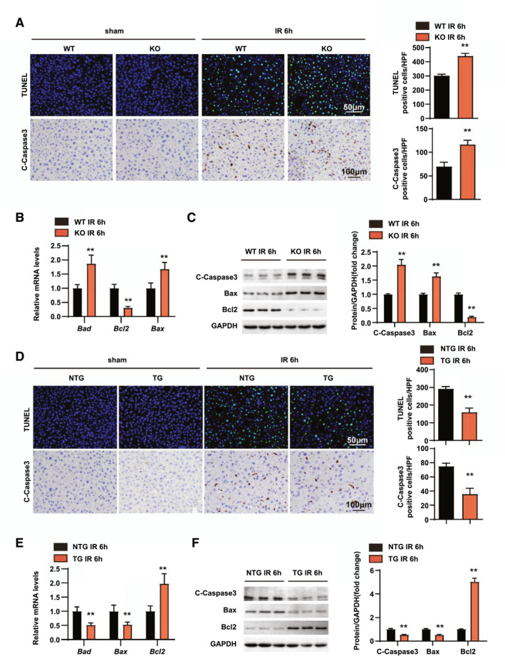
RGS14 alleviates apoptosis in hepatic IRI. (A) TUNEL staining and statistics in ischemic liver sections from mice in the indicated groups (n = 4/group). Immunohistochemical staining of cleaved‐caspase‐3 expression and statistics in ischemic liver sections from mice in the indicated groups (n = 6/group). (B) Relative mRNA expression levels of Bad, Bcl2, and Bax in liver tissues of WT and RGS14‐KO mice at 6 h after IR (n = 4/group). (C) Western blot analysis of cleaved‐caspase‐3, Bax, and Bcl2 expression in livers from WT and RGS14‐KO mice at 6 h after IR (n = 3/group). (D) TUNEL staining and statistics in ischemic liver sections from mice in the indicated groups (n = 4/group). Immunohistochemical staining of cleaved‐caspase‐3 expression and statistics in ischemic liver sections from mice in the indicated groups (n = 6/group). (E) Relative mRNA expression levels of Bad, Bcl2, and Bax in liver tissues of NTG and RGS14‐TG mice at 6 h after IR (n = 4/group). (F) Western blot analysis of cleaved‐caspase‐3, Bax, and Bcl2 expression in livers from NTG and RGS14‐TG mice at 6 h after IR (n = 3/group). GAPDH served as a loading control. All data are shown as the mean ± SD. *p < 0.05, **p < 0.01. C‐Caspase3, cleaved caspase‐3; HPF, high‐power field