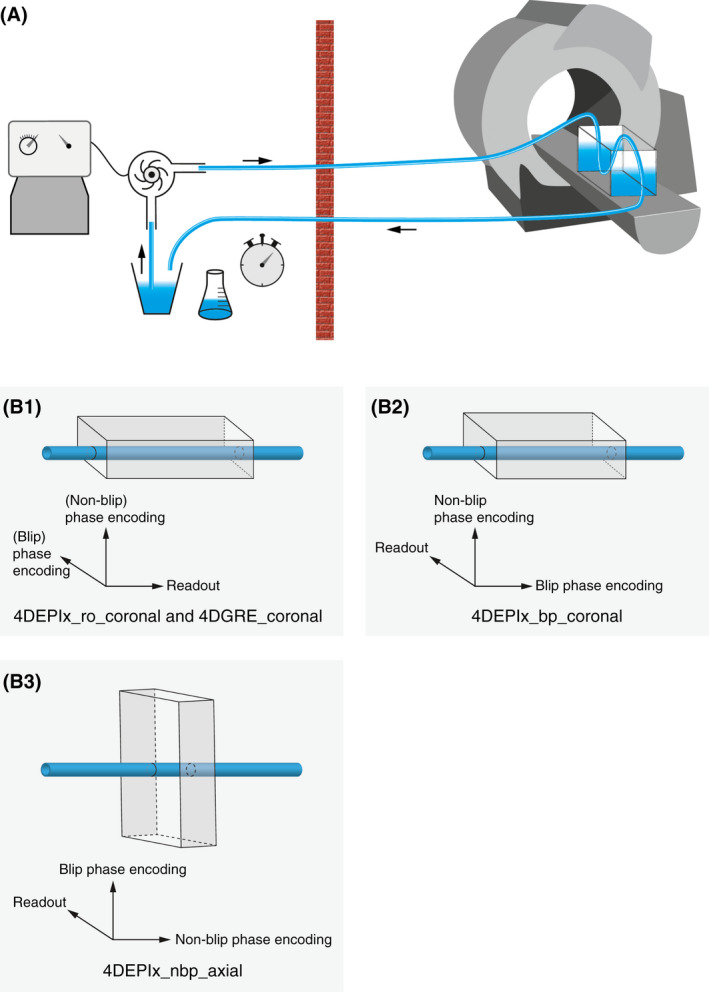
FIGURE 1.

(A) Phantom setup for experiment 1. A tank with straight tube phantom is positioned inside the MRI scanner. A pump outside the scanner room maintains steady flow, which is calibrated by timed volumetric collection at the returning tube. (B) The orientations of the 4D flow MRI acquisitions with respect to the main flow direction in the tube. In (1), 4DEPI5_ro_coronal refers to the coronally oriented EPI acquisition with the readout gradient aligned parallel to the main flow direction; in (2), 4DEPI5_bp refers to the coronally oriented EPI acquisition with the blip phase‐encoding gradient aligned parallel to the main flow direction; and in (3), 4DEPI5_nbp_axial refers to the axially oriented EPI acquisition with the readout and blip phase‐encoding gradient both perpendicular to the main flow direction. The 4D gradient echo (4DGRE) has the same orientation as 4DEPI5_ro_coronal. The schematically drawn scan volumes are not up to scale with the phantom dimensions
