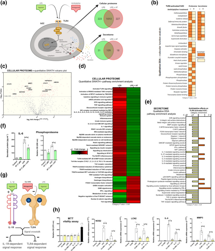
FIGURE 2.

Amitriptyline (AT) reverts TLR4‐mediated proteomic changes in human osteoarthritis (OA) chondrocytes (hOCs). All experiments were independent, normalised by control and expressed as the mean ± SEM and statistically analysed by non‐parametric Krustal‐Wallis test coupled with Dunn's post‐test (* P<0.05). hOCs were stimulated by LPS (100 ng·ml−1) and cotreated with AT (1 μM) for 24 h. (a) Diagram of TLR4 signalling inhibition and proteome number identification in qualitative analysis (DDA) data (n = 3). (b) Qualitative data (DDA) clustering into molecular function categories from TLR4‐activated hOC proteome (n = 3). (c) Quantitative data (SWATH) shown as a volcano plot (P value vs. fold change) of protein expression changes by AT in TLR4‐activated hOCs (n = 3). (d) Quantitative data (SWATH) pathway enrichment analysis of AT effects on TLR4‐activated hOCs (n = 3). (e) Qualitative secretome data (DDA) pathway enrichment analysis of AT effects on TLR4‐activated hOCs (n = 3). (f) Secreted levels of protein IL‐6 (pg·ml−1) (elisa) and relative OD of malachite green total phosphoproteome (n = 3). (g) Diagram of TLR4 and IL‐1 receptor ® crosstalk in the context of CLI‐095 inhibition in hOCs. (h) MTT cell viability assay and innate immune response factor mRNA expression (RT‐PCR) were determined in hOCs pretreated with CLI‐095 (1 μM) for 3 h before TLR4 or IL‐1R (IL‐1β [0.1 ng·ml−1]) activation for 24 h (n = 6)
