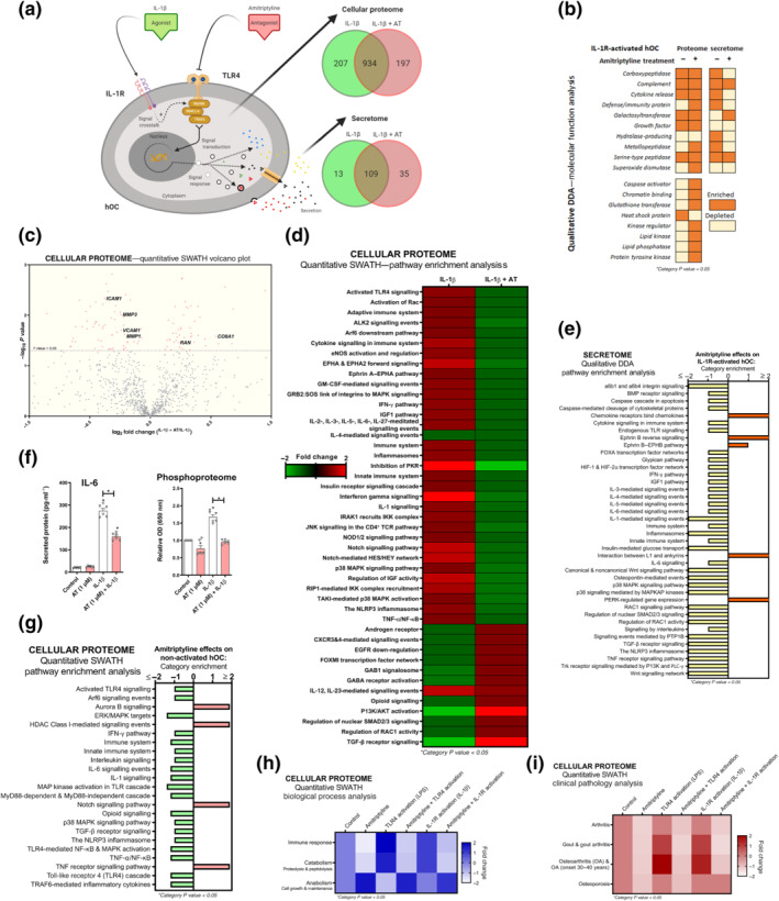
FIGURE 4.

Amitriptyline (AT) reverts IL‐1 receptor (R)‐mediated proteomic changes in human OA chondrocytes (hOCs). All experiments were independent, (n=6) normalised by control and expressed as the mean ± SEM and statistically analysed by non‐parametric Krustal‐Wallis test coupled with Dunn's post‐test (* P<0.05). hOCs were stimulated with IL‐1β (0.1 ng·ml−1) and cotreated with AT (1 μM) for 24 h. (a) Diagram of TLR4 signalling inhibition and qualitative data (DDA) protein number identification from IL‐1 receptor (R)‐activated hOC proteome (n = 3). (b) Qualitative data (DDA) clustering into molecular function categories from IL‐1 receptor‐activated hOC proteome (n = 3). (c) Quantitative data (SWATH) of protein expression changes by AT shown as a volcano plot (P value vs. fold change) from IL‐1 receptor‐activated hOC proteome (n = 3). (d) Quantitative data (SWATH) pathway enrichment analysis of AT effects on IL‐1 receptor‐activated hOCs (n = 3). (e) Qualitative secretome data (DDA) pathway enrichment analysis of AT effects on IL‐1receptor‐activated hOCs (n = 3). (f) Secreted levels of protein IL‐6 (pg·ml−1) (elisa) and relative OD of malachite green total phosphoproteome (n = 3). (g) Amitriptyline effects on pathway enrichment of non‐activated hOCs using quantitative (SWATH) proteome data (n = 3). (h) Quantitative data (SWATH) clustering into biological processes categories from IL‐1 receptor‐activated hOC proteome (n = 3). (i) On clinical pathology analysis using quantitative data (SWATH) of AT effects on stimulated and non‐stimulated hOCs (n = 3)
