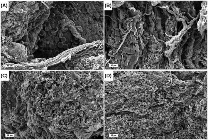
FIGURE 2.

Representative SEM images from eight prospectively collected portal vein thrombus samples. (A,B) Collagen bundles and some biconcave‐shaped red blood cells. Fibrin is focally present. (C,D) Fibrin networks with mostly biconcave‐shaped red blood cells
