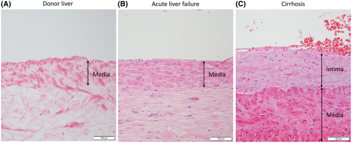
FIGURE 3.

Representative images of the portal vein wall in H&E‐stained sections from hilar liver tissue samples from human donor livers, patients with acute liver failure, and patients with cirrhosis without PVT. The tunica intima at the liver hilum in human donor liver and patients with acute liver failure consists of a flat lining of endothelial cells and is almost unrecognizable, and therefore not measurable (A,B). The portal vein vessel wall at the liver hilum in patients with cirrhosis without PVT shows a thickened tunica intima (C) [Color figure can be viewed at wileyonlinelibrary.com]
