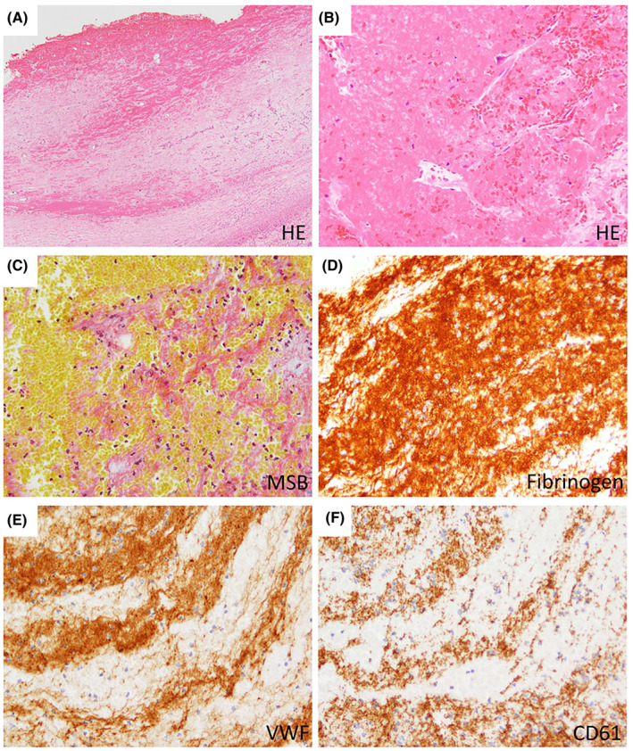
FIGURE 5.

Histopathology of PVT at the hilar region of explanted livers. Representative examples of intimal fibrosis with fibrin thrombus of the portal vein. A fibrin thrombus overlying the thickened intima is observed (A; ×20). The fibrin thrombus consists of aggregated eosinophilic materials and blood contents (B; ×200), and these parts are stained orange to red by MSB, indicating fibrin (C; ×200). The fibrin thrombus is immunoreactive to fibrin/fibrinogen, VWF, and CD61; and fibrin/fibrinogen is most widely positive (D–F; ×200) [Color figure can be viewed at wileyonlinelibrary.com]
