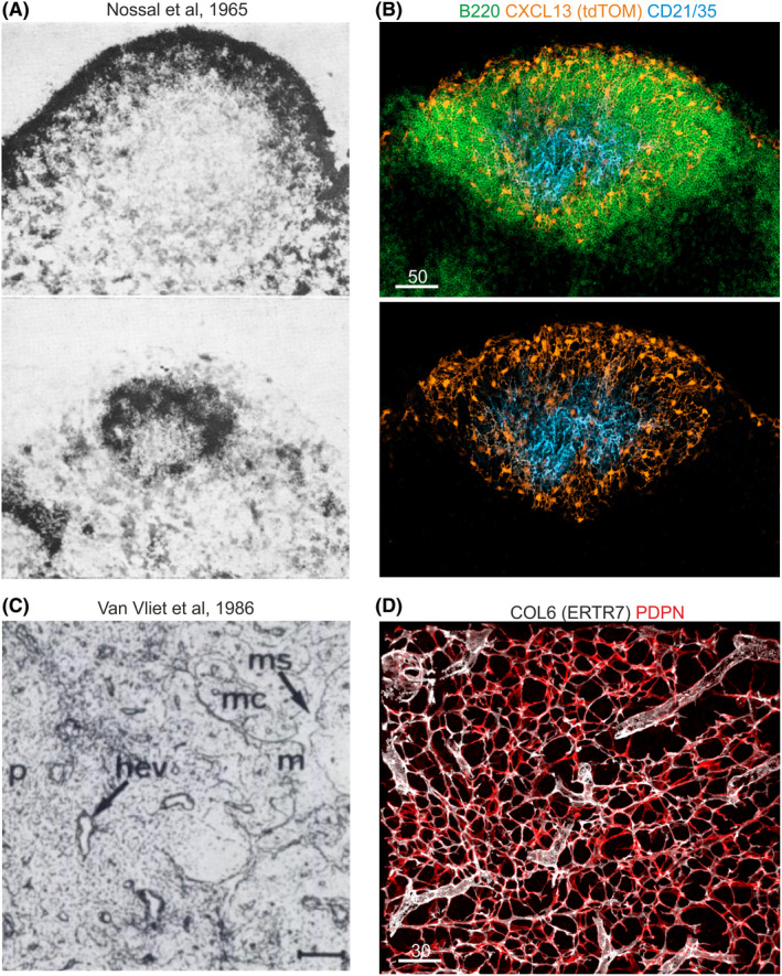
FIGURE 1.

Fibroblastic reticular cell networks. (A) Nossal et al 17 have analyzed the retention of flagellar antigens labeled with radioactive Iodine (125I) in primary and secondary follicles of rat lymph nodes. Scintillation counting and autoradiography highlighted a reticular cell network, which retains antigen early in perifollicular (upper image) and later in the center of the follicle (lower image). Reprinted with the kind permission from Immunology (John Wiley & Sons). (B) Cxcl13 promoter activity revealed by the Cxcl13‐Cre/tdTomato model as fluorescent staining in B cell zone reticular cell networks that underpin subcapsular and perifollicular regions of mouse lymph nodes. Central follicles exhibit follicular dendritic cells (FDCs), which express the Cxcl13‐tdTomato transgene and complement receptors 1 and 2 (CD21/35). (C) Staining of mouse lymph node fibroblasts by Van Vliet et al 24 using the ER‐TR7 antibody that binds to the extracellular matrix component collagen type VI and stains the reticular fibroblast network in paracortical (p) and medullary (m) regions, surrounding medullary sinuses (ms) and within medullary chords (mc). Reprinted with the kind permission from The Journal of Histochemistry and Cytochemistry (SAGE Publications). (D) ER‐TR7 staining of the murine lymph node T cell zone marks the fibrillar core of the microfluidic conduit network, which is produced by podoplanin (PDPN)‐expressing T cell zone reticular cells
