
Fig. 1: Cardiomyocytes (CMs) adopt a distinct nuclear architecture during development with high amounts of peripheral chromatin.
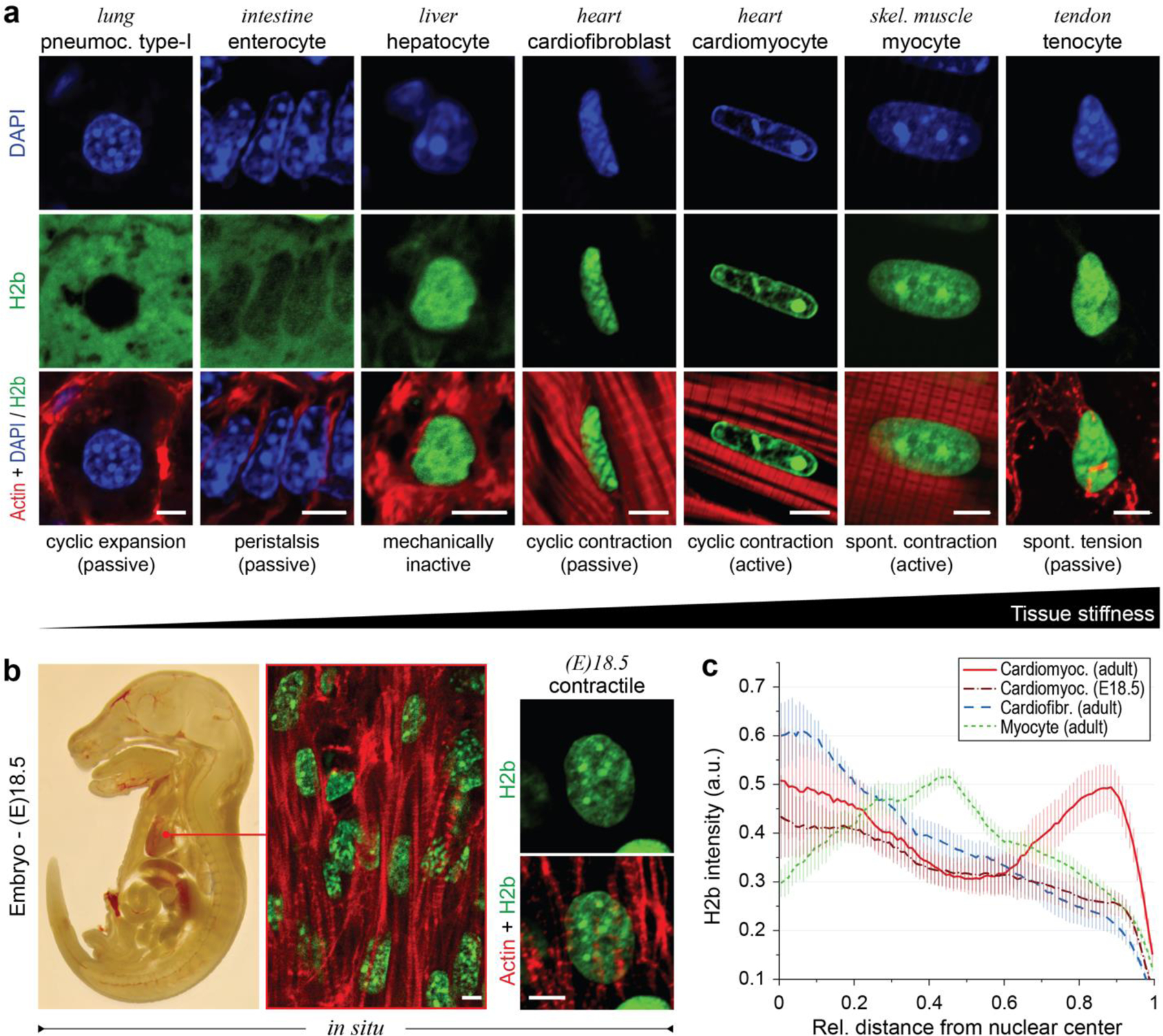
a) Tissues with diverse mechanical characteristics were harvested from adult H2b-eGFP mice and stained for actin. DAPI was used as DNA counterstain for soft tissues with weak GFP fluorescence. b) H2b-eGFP mice embryos were harvested at day 18.5, sectioned and stained for actin. Left: whole embryo mid-section. Middle: close-up of embryonic heart. Right: close-up of an embryonic CM nucleus, which showed a diffuse nuclear organization unlike adult CMs in (a). c) H2b intensity was analyzed with respect to its relative distance to the nuclear center (1=periphery) in nuclei of different cell types in situ. Adult CMs showed a high ratio of peripheral chromatin compared to other cell types. n=5; all scales=5 µm.