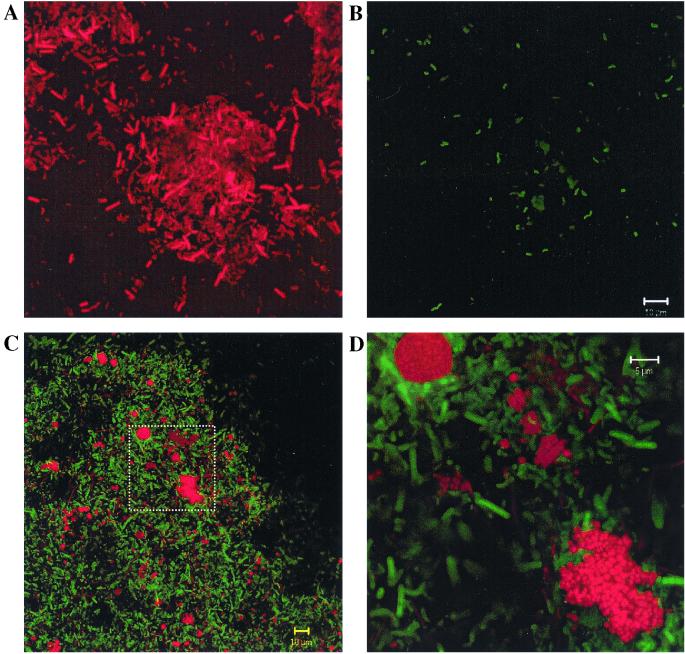
FIG. 3.
Confocal laser scanning micrographs of FISH-probed ABR samples. (A and B) Cells in the same field. Cells were detected with probe EUB338 (A) and with probe DSV698 (B). The vibrio morphology typical of Desulfovibrio spp. is exhibited by several cells in panel B. (C and D) Cells in a sludge floc detected with probes ARC915 (red) and EUB338 (green). The rectangle in panel C is magnified in panel D. In this image, the cells detected with the ARC915 probe appear more clearly as microcolonies of coccoid cells.

