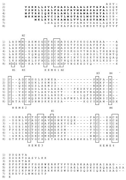
FIG. 1.
Comparison of the sequences of ST cyt's and the tetraheme domains or subunits of FL cyt's from different species. Bacterial strain H1R ST cyt (2) (rows 1), S. oneidensis MR1 ST cyt (rows 2), S. frigidimarina NCIMB400 ST cyt (EMBL accession no. AJ000006) (rows 3), S. oneidensis MR1 FL cyt (rows 4), S. frigidimarina NCIMB400 FL cyt (33) (rows 5), S. frigidimarina NCIMB400 IfcA cytochrome (7) (rows 6), W. succinogenes (36) (rows 7), and D. vulgaris Miyazaki cytochrome c3 (12) (rows 8) sequences are shown. The heme binding sites and sixth heme ligand histidines are boxed. The sixth ligands to Shewanella hemes 1 to 4 are labeled H1 to H4 above the sequence and those for the Desulfovibrio hemes are labeled H1 to H4 below the sequence. To conserve space, no attempt to precisely align the N and C termini was made. Gaps introduced to optimize alignment are indicated by the dashes.

