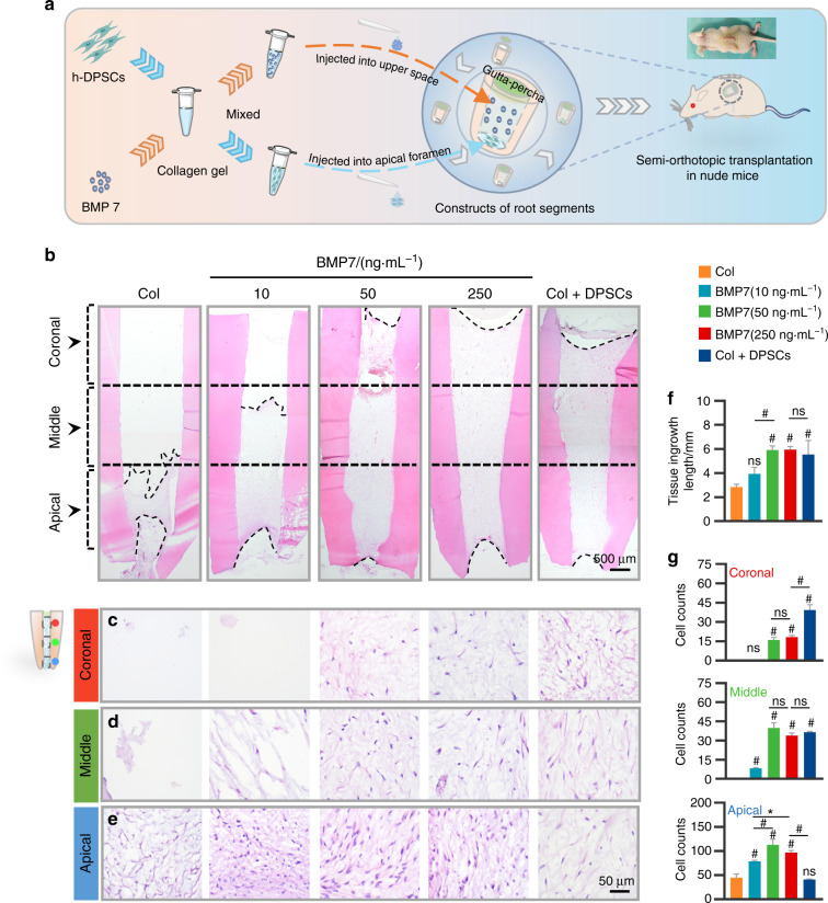
Fig. 6.
The evaluation of ingrowth of pulp tissue in the nude mice model. a Schematic diagram of the ectopic pulp regeneration model in nude mice. Collagen gel pre-mixed with h-DPSCs or BMP7 was respectively injected into the upper space and the apical foramen of the RSs, which were subcutaneously transplanted into the back of nude mice for 6 weeks. b The overall view of pulp-like tissue regeneration in the RSs by H&E staining. The distribution of the migrated cells was evaluated in the coronal (c), middle (d), and apical (e) regions in the root canal. f Quantification of the tissue ingrowth length in b. g Quantification of the average cell number per field in the coronal, middle, and apical regions. *P < 0.05, #P < 0.01 vs the Col group. NS, no significance. Scale bar: b 500 μm, c–e 50 μm

