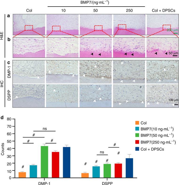
Fig. 7.
The evaluation of the dentin-pulp-like tissue regeneration in vivo by H&E and IHC. a, b The regenerated dentin-pulp-like tissues showed by H&E staining. b showed the enlarged images of the framed area in a. The black arrowheads showed polarized odontoblasts-like cells layer in the BMP7(50 ng·mL−1), BMP7(250 ng·mL−1), and Col + DPSCs groups. c IHC showed the DMP-1-positive or DSPP-positive odontoblasts-like cells (white arrowheads). d Quantitative analysis of the DMP-1-positive or DSPP-positive odontoblasts-like cell number. *P < 0.05. #P < 0.01 vs. the Col group. NS, no significance. Scale bar: a 200 μm, b 50 μm, c 100 μm

