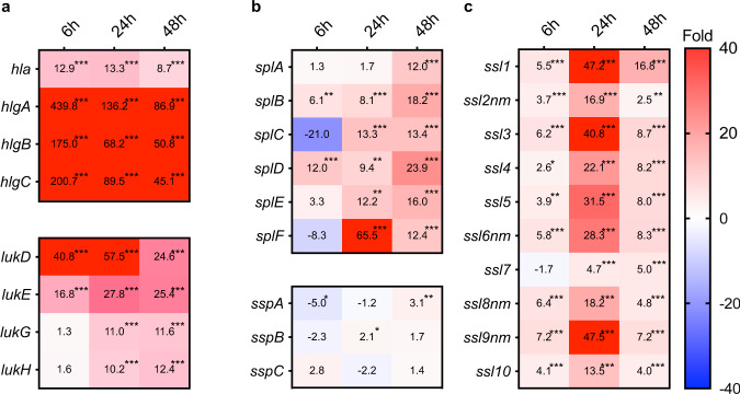
Fig. 5. Time course of expression changes in virulence factors in S. aureus-infected liver compared with culture medium.
Time course of expression changes for toxins (a), proteases (b), and superantigens (c). Values in the boxes show fold-change compared with S. aureus cultured in the TSB medium and the boxes filled with red indicate >40-fold change. The asterisk *. **, and *** indicate FDR p-values < 0.05, <0.01, and <0.001, respectively. lukG and lukH genes in (a) are annotated as lukS and lukF in Refseq (NZ_CP023390.1), although we annotated these as lukGH genes as these genes are not related to the PVL phage and are similar to the lukGH gene.

