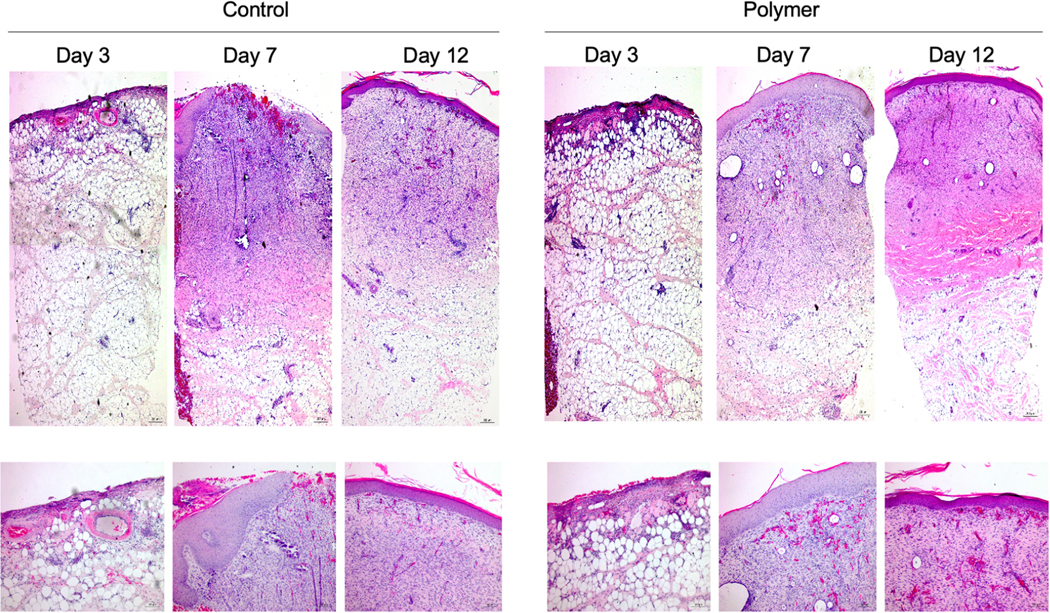
Fig. 9 –
In Animal 3 in the most cranial wounds, H&E staining revealed similar histo-architecture between control vs. polymer groups in FT wounds in the biopsies taken from interstices. Biopsies were taken at Days 3, 7, and 12 and were formalin fixed and paraffin embedded. Sections were then stained for H&E and images were captured for the control group (A) and polymer (B) at 5X magnification (top) and 10X magnification (bottom).

