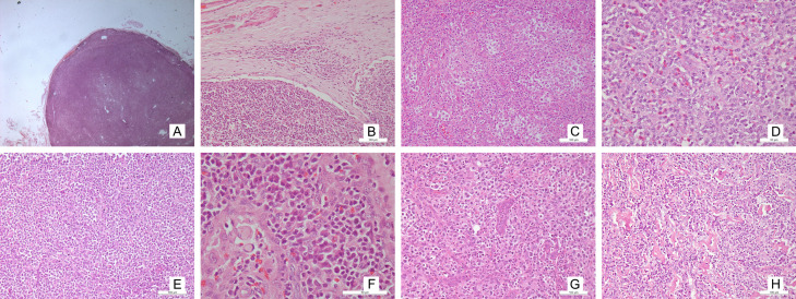
Figure 4.
Microphotograph shows: Diffuse effacement of architecture with paracortical expansion [A: H&E ×16] Presence of patent marginal sinus [B: H&E ×100] Intermediate sized atypical lymphoid cells having clear cytoplasm [C: H&E ×100] Polymorphous population with predominant eosinophil population [D: H&E x100] AITL with predominant lymphocyte population [E: H&E ×200] Polymorphous population with predominant plasma cells and Russel bodies [F: H&E ×100] Increased high endothelial venules [G: H&E ×100]. Eosinophilic hyaline material deposition [H: H&E ×100].

