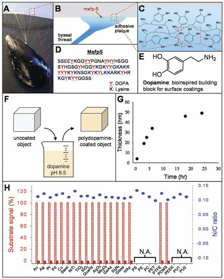
FIGURE 1.
The formation of PDA coatings onto plenty of materials’ surfaces. (A) Photograph of a mussel attached to PTFE. (B,C) Interfacial location of Mefp-5, as well as the representation of catechol and amine groups. (D) The sequence of amino acid in Mefp-5. (E) DA contains same functional catechol and amine groups. (F) A schematic illustration of PDA deposition. (G) Thickness of PDA coating. (H) XPS evaluation of PDA-coated surfaces (Lee et al., 2007). Copyright 2007, The American Association for the Advancement of Science.

