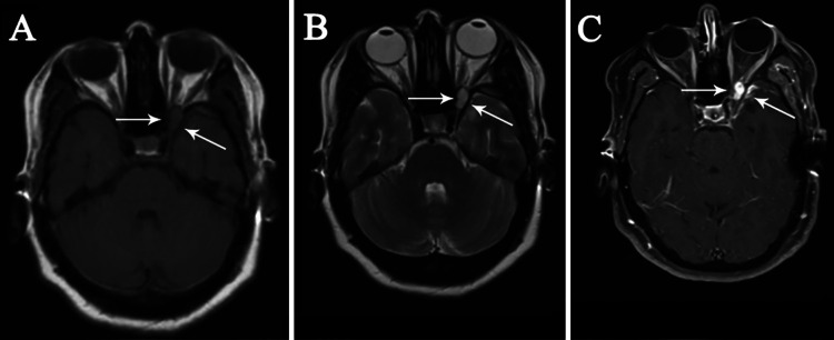
FIG. 2.
Preoperative axial T1-weighted (A), T2-weighted (B), and contrast-enhanced (C) MRI sequences showing an extradural, well-circumscribed round lesion as isointense on T1-weighted and hyperintense on T2-weighted images. T1-weighted images with gadolinium-DTPA demonstrate a mass with avid enhancement compressing the optic nerve. The dura overlying the ACP is also enhancing (small white arrows).

