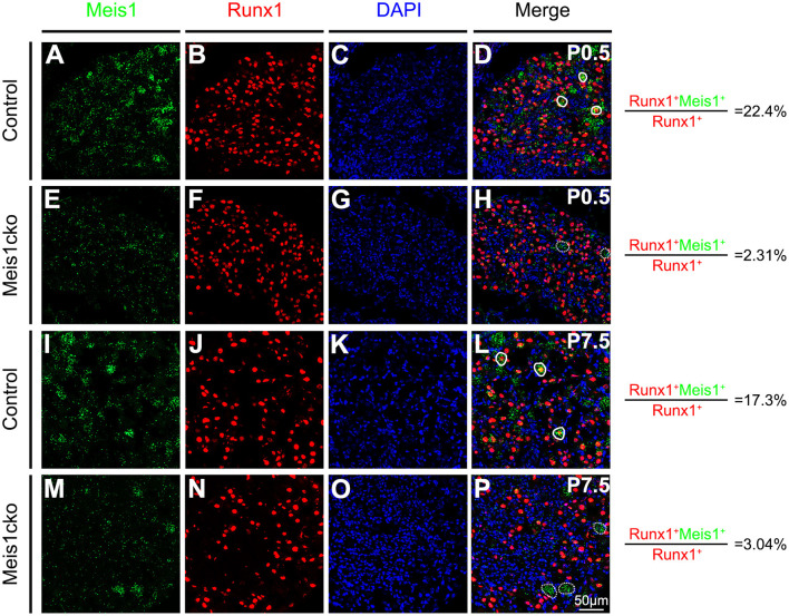
Figure 2.
Comparing Meis1 expression between Meis1cko and control DRGs. Representative images of lumbar DRG sections costained with an antibody against the Runx1 protein (red) and Meis1 mRNA (green) in Meis1cko and control mice at P0.5 and P7.5. (A–H) 22.4% (684/3052 in control mice) and 2.31% (42/1818 in Meis1cko mice) of Runx1+ neurons coexpressed Meis1 at P0.5. (I–P) A total of 17.3% (370/2142 in control mice) and 3.04% (33/1086 in Meis1cko mice) of Runx1+ neurons coexpressed Meis1 at P7.5. Solid lines indicate Meis1+ neurons coexpressing Runx1. Meis1 was expressed in large neurons indicated with a dotted line. DAPI-stained cell nuclei appeared blue. Quantitative data are shown to the right of the panels. Scale bar: 50 μm.

