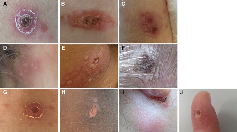
Figure 2.
Photographs of the inoculation sites of 10 patients with ulceroglandular tularemia. Panels (C), (D), and (E) show clustered vesicles, pustules, and ulcers; in panels (A), (B), (F), (G), and (H), multiple distinct lesions surrounding the major ulcer or eschar can be seen. Panel (J) depicts the site of a wood mouse bite, from which F. tularensis was cultivated.

