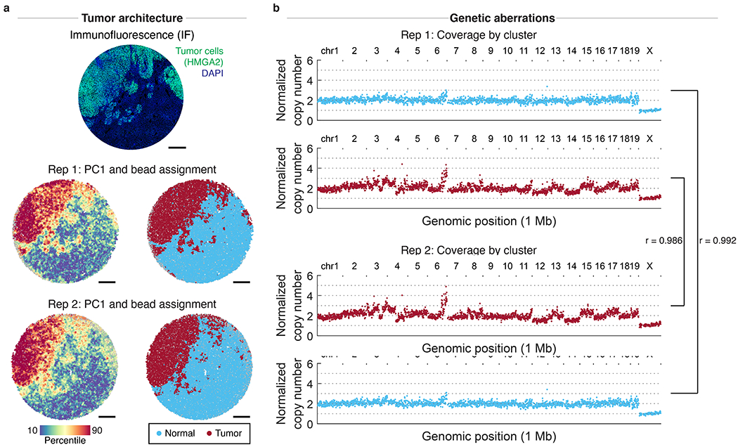
Extended Data Figure 9: Reproducibility of slide-DNA-seq across serial sections.

a, Immunofluorescence (IF) against tumor marker HMGA2 (top) and two slide-DNA-seq replicates (center, bottom) were performed on two serial sections of a mouse liver metastasis. Beads colored by PC1 scores (left) and cluster assignment (right) show similar spatial architecture between replicates. Scale bars, 500 μm. b, Aggregate copy number profiles of normal and tumor beads show high correlation (Pearson’s r = 0.986 and 0.992) between the two replicates.
