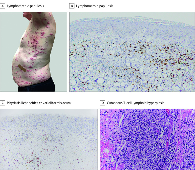
Figure. Clinical Images and Histologic Findings Consistent With Lymphomatoid Papulosis, Pityriasis Lichenoides et Varioliformis Acuta, and Cutaneous Lymphoid Hyperplasia.
A, Generalized eruption of hemorrhagic-necrotic papules and plaques. B, Infiltrate composed of CD30-positive atypical cells (original magnification ×20). C, Infiltrate with scattered CD30 expression (original magnification ×10). D, Dense infiltrate composed of medium to large, immunoblastic and pleomorphic lymphocytes, with well-visualized atypia (hematoxylin-eosin stain; original magnification ×40).

