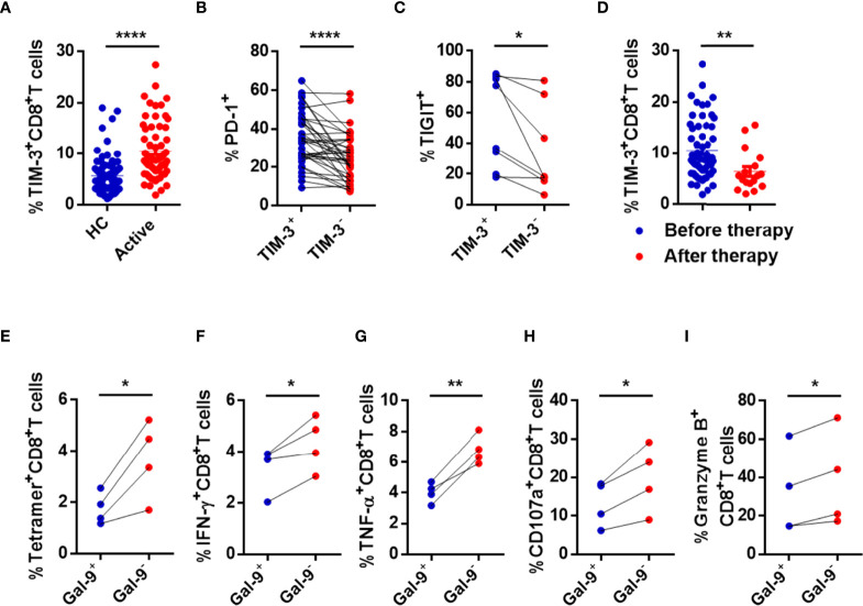
Figure 4.
CD8+ T cells are immune exhausted through the Gal-9/TIM-3 axis in active CHB patients. (A) Comparison of the levels of TIM-3 expressed in circulating CD8+ T cells in active CHB patients and HCs. (B) The percentages of PD-1 expressed in TIM-3+CD8+ T cells and TIM-3-CD8+ T cells in peripheral blood from active CHB patients. (C) The percentages of TIGIT expressed on TIM-3+CD8+ T cells and TIM-3-CD8+ T cells in peripheral blood from active CHB patients. (D) The percentage of TIM-3 expressed on CD8+ T cells in peripheral blood from active CHB patients with or without antiviral treatment, respectively. (E–I) Gal-9+ NK, Gal-9- NK, and CD8+ T cells were sorted from active CHB patients, cocultured in vitro, and stimulated with anti-CD3/anti-CD28 for 3 days. The frequency of tetramer+CD8+ T cells (E) and the production of the cytokines IFN-γ, TNF-α, CD107a, and granzyme B (F-I) by CD8+ T cells were analyzed. Results are expressed as the mean ± SEM, and the number of samples (n) in each group was ≥ 3. Unpaired t-test was used to compare two independent groups. A paired t-test was used to compare paired samples. *P < 0.05; **P < 0.01; ****P < 0.0001.

