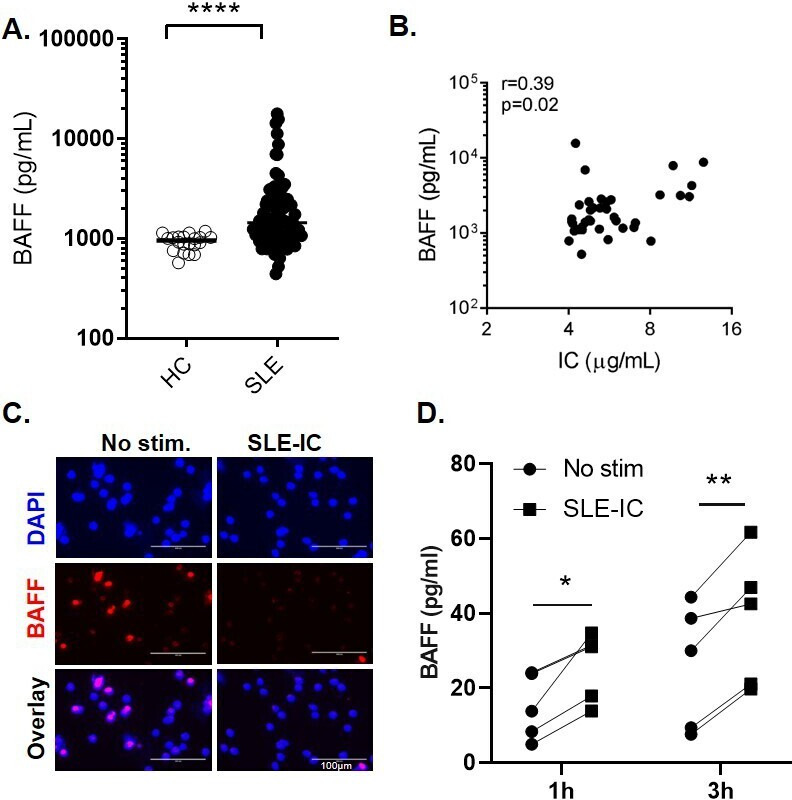
Figure 1.

SLE immune complexes induce the release of BAFF from neutrophils. (A) Serum BAFF levels in patients with SLE (n=60) and healthy controls (HC) (n=20) measured by ELISA. Each symbol represents an individual; horizontal lines indicate mean. (B) Correlations between serum BAFF levels and circulating immune complexes (IC). (C–D) Neutrophils isolated from healthy controls (HC) were left untreated or stimulated with SLE-IC for 1 or 3 hours. (C) Immunofluorescent microscopic imaging showing nuclear DNA (blue) and BAFF (red) of neutrophils stimulated with SLE-IC for 1 hour. Data are representative of three independent experiments. Scale=100 µm. (D) BAFF release in cell supernatants in neutrophils from 5 individual HC (1 or 3 hours of cell culture), determined by ELISA. *p<0.05, **p<0.01 and ****p<0.0001, significance determined by Mann-Whitney U test or Student’s paired t-test, correlations determined by Spearman’s correlation.
