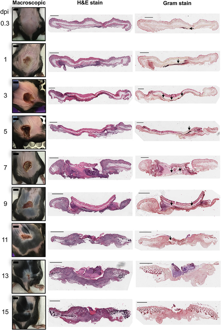
FIG 2.
Tissue damage and bacterial localization. Representative photographs of lesions (macroscopic) throughout the infection, H&E staining of histology slides, and modified Gram staining of tissue are shown. Bars on macroscopic mouse pictures, 0.5 cm. Sections are 5 μm thick. Bars on tissue sections, 1 mm. Arrows indicate S. aureus.

