FIG 6.
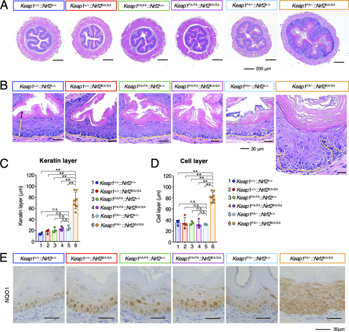
Hyperkeratosis and hyperplasia of the esophagus in Keap1FA/–::Nrf2SA/SA mice. (A, B) HE staining of mouse esophageal cross sections. The yellow and black double-edged arrows in the left panel show the epithelial cell and keratinous layers, respectively. The yellow dotted lines show the boundary of the epithelial cell layer. Hyperkeratosis and hyperplasia of the esophagus are specifically induced in Keap1FA/–::Nrf2SA/SA mice. (C, D) Thickness of keratinous layers (C) and cell layers (D) of the esophageal epithelium. The bar graphs show the means ± SD. **, P < 0.01 by one-way ANOVA with Tukey’s multiple-comparison test. (E) Immunohistochemistry of NQO1.

