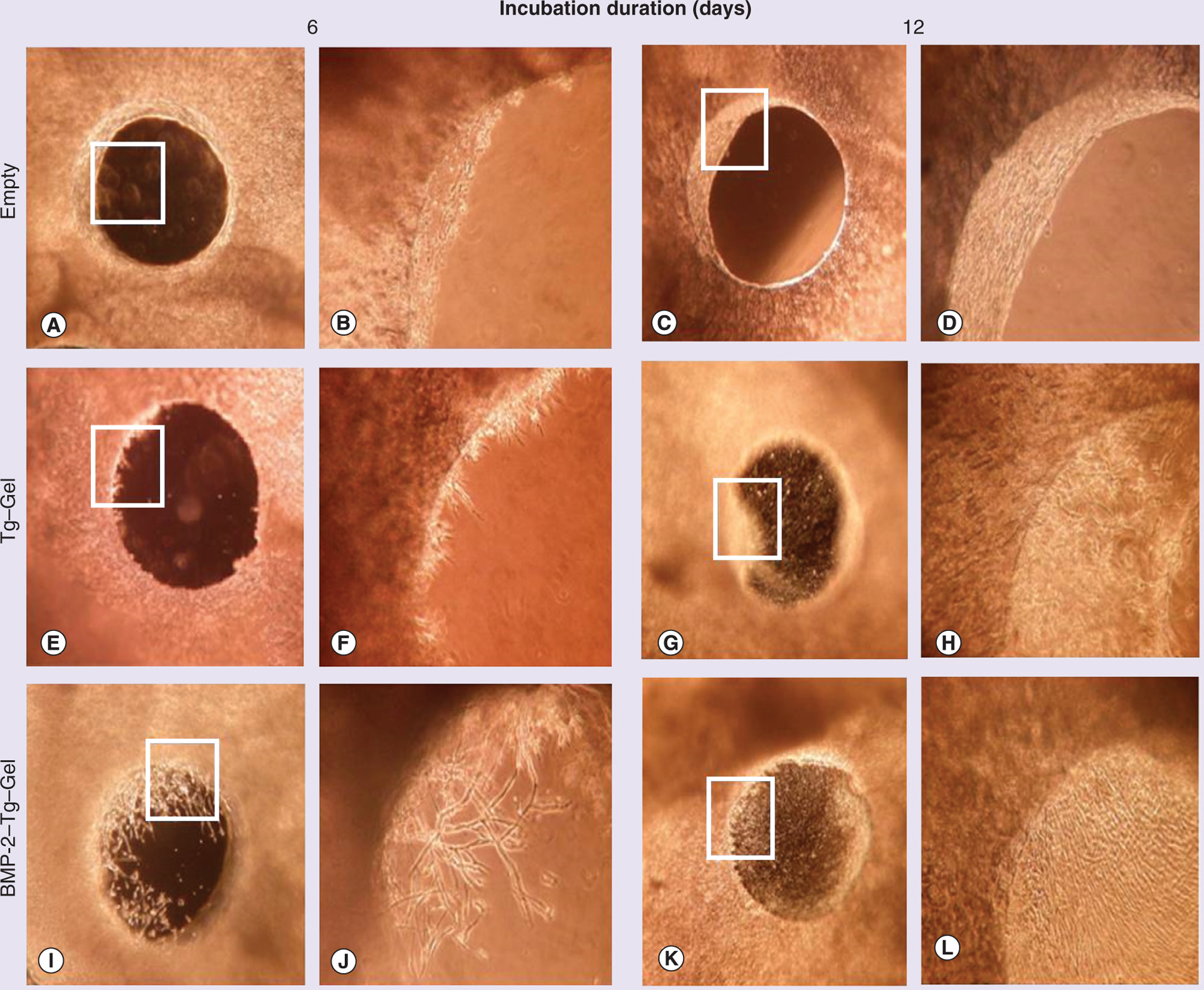
Figure 4. Bone repair through gel constructs in calvaria bone organ culture model.

Phase contrast images of 1.0-mm defects on days 6 and 12 of various treatments. Empty served as a negative control without any gel. A total of 20 μl of assigned gel construct was applied to each calvaria defect placed at the bottom of a 24-well cell culture plate accordingly. Images (A, C, E, G, I & K) were visualized at a magnification of 40×, and images (B, D, F, H, J & L) were pictured at a magnification of 100× using an inverted light microscope (Nikon, Japan), and are higher magnification images of the boxed sections in (A, C, E, G, I & K), respectively. BMP-2–Tg–Gel: Transglutaminase-crosslinked gelatin gel with BMP-2; Tg–Gel: Transglutaminase-crosslinked gelatin gel.
