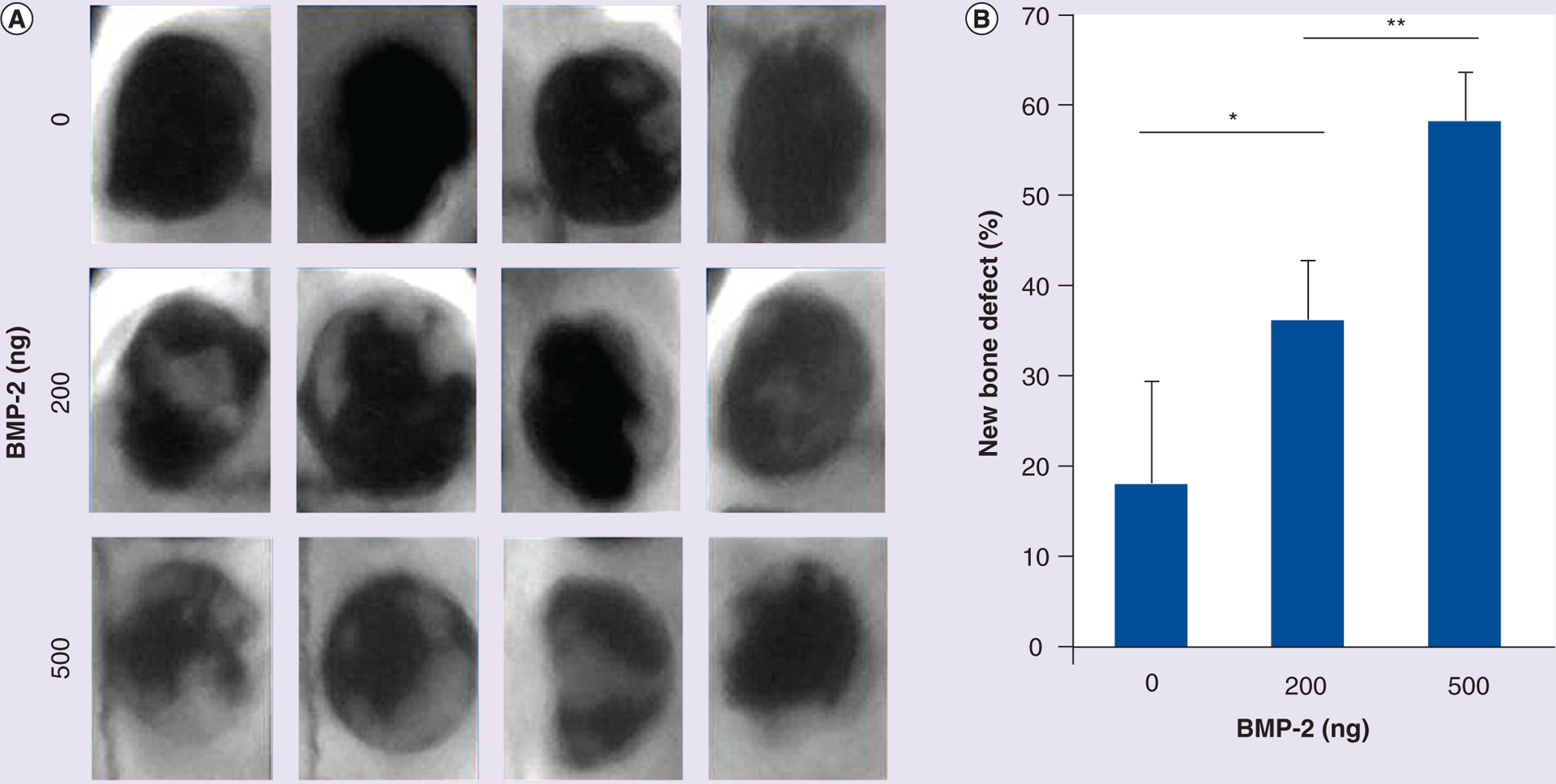
Figure 6. Bone repair in cranial defect models.

After 14 days of subcutaneous injection, x-ray images of calvaria bone repair were captured for the 12 rat models. (A) Faxitron images of bone repair at the defect site 14 days after the administration of tethered BMP-2 (0, 200 and 500 ng). (B) New bone area was analyzed and calculated using ImageJ software (NIH, MD, USA). Data are expressed as mean ± standard deviation.
*p < 0.05; **p < 0.01.
