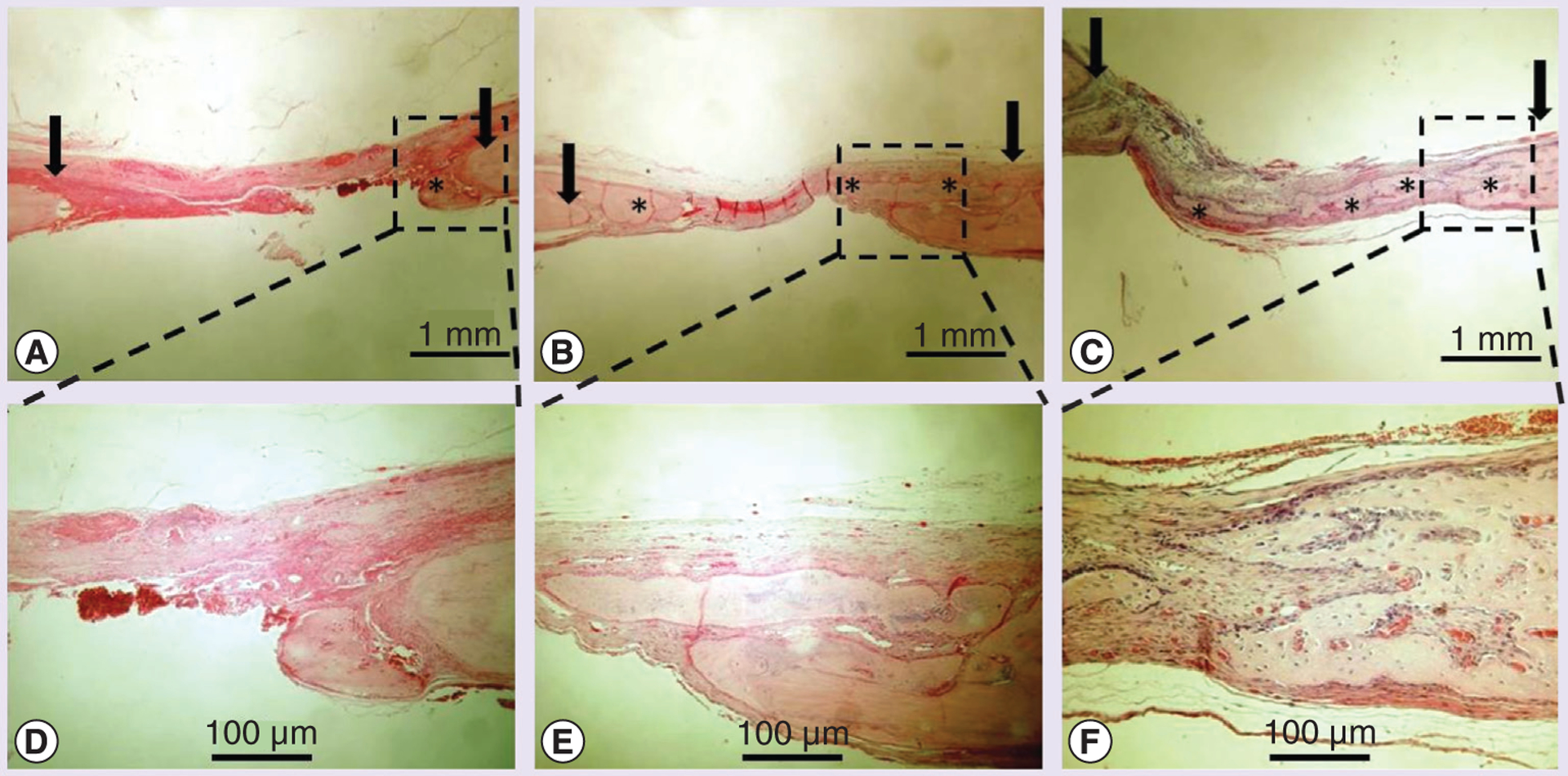
Figure 7. Osteoinductivity of transglutaminase-crosslinked gelatin gel with BMP-2 in vivo cranial defect models.

A total of 14 days after grafting, explants underwent histology evaluation (arrow indicates bone defect edge, * indicates new bone formation). Hematoxylin and eosin staining was performed for group (A) transglutaminase-crosslinked gelatin gel (Tg–Gel), (B) Tg–Gel + 200 ng BMP-2 and (C) Tg–Gel + 500 ng BMP-2 (magnification: 20×). Bone formation and tissue reorganization in the boxed areas of (A–C) are shown with higher magnification in (D–F), respectively.
